
Mummy CT scans provide fascinating insights on ancient Egyptian life.
CT scans indicate the mummies.

The purpose of CT scanners is to diagnose the living by precisely mapping concealed tumours, obstructed arteries, and fractured bones. Restoring individual stories that have been silent for thousands of years is an unanticipated outcome of applying the same technology to the ancient past.
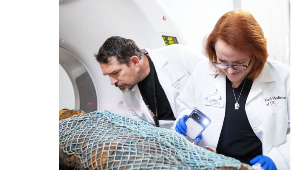
Two ancient Egyptian males were brought into a Los Angeles medical imaging suite more than 2,200 years after they had passed away. They weren't patients. They did not ride gurneys to get there.
The soldiers arrived in their coffins sealed, with preserved records of their daily life, physical hardships, and state of health.
CT scans indicate the mummies.
Priests from Egypt were the two males. Nes-Min, who lived in 330 BCE, was one among them. Nes-Hor, who lived in 190 BCE, was the other.
Because to their meticulous preservation, both bodies had withstood centuries of handling, wars, and empires. But beneath the layers of cloth and glue, many aspects of their lives were still obscured.
When radiologists from USC's Keck Medicine put each corpse onto a CT scanner while it was still in the lower half of its sarcophagus, that changed everything. The weight of each casket was around 200 pounds. Not a single wrap was disturbed during the scans, which caught their complete bodies.
Even experienced professionals were taken aback by what the scans showed. Fine details, including the lower lips and eyelids, were revealed by a cutting-edge 320-slice CT scanner.
Such subtleties helped the guys feel less like artefacts in a museum and more like humans who previously lived, worked, and grew old.
Health issues that seem familiar
The scans demonstrated how both priests had been impacted by time in ways that viewers of today may recognise. There were indications of a collapsed lower back vertebra in Nes-Min, the older mummy.
Wear and tear and natural ageing were probably the causes of the damage. To put it simply, he most likely suffered from lower back pain.
He was interred among a number of artefacts, such as a fish and scarab beetles. There were strands of colourful beads around his neck and a thickly beaded net across his torso. Although his rank and opinions were implied by these objects, his bodily discomfort was subtly conveyed by the CT pictures.
The body of Nes-Hor revealed a different tale. His scans showed a badly degraded hip and dental issues. According to the damage, he was older than Nes-Min when he passed away. These discoveries reminded viewers that everyday physical problems are nothing new and put aches, injuries, and chronic agony firmly in the past.
Better responses and new technology
Following the preliminary results, the research went beyond simple imaging. The scans and analysis were directed by Summer Decker, Ph.D., director of the USC Center for Innovation in Medical Visualisation and head of 3D imaging at Keck Medicine.
"The team's experience and Keck Medicine's access to the newest high-level scanning technology enable these scans to yield a wealth of information."
Although the mummies had already been scanned, new developments in scanning technology have yielded results that are significantly more thorough and detailed. The high-resolution photos gave researchers a better understanding of their lives by revealing details that had not been known before.
Transforming CT scans become history that can be touched
The team created intricate 3D computer models of the two guys using the CT data. They created life-size 3D prints of hips, skulls, spines, and a number of the artefacts interred with Nes-Min using those models. The printers were of the same type used in medical settings.
For a long time, mummies have been a mystery. According to anthropologist Diane Perlov, Ph.D., senior vice president for special programs at the California Science Center, "seeing beneath the surface to reveal the specific lived experience of individuals is incredibly exciting."
"We have a powerful window into the world of ancient people and past civilisations that might otherwise be lost thanks to modern scientific technology."
Teachings from CT images of mummies
At Keck Medicine, the same instruments used on the mummies are utilised in routine medical procedures. Hundreds of cross-sectional images, also referred to as "slices," are produced by CT and MRI scans. These photos are digitally stacked by experts to produce intricate 3D reconstructions of bones and organs.
It is possible to measure, study, or print those models. They help surgeons comprehend intricate anatomy prior to surgery. Both handling and, in some situations, implanting the printed parts are safe.
"Clinicians like surgeons can accurately measure hard-to-detect tumours, examine the intricate structure of a patient's heart or liver, or determine how best to repair a shoulder or hip through 3D visualisation, modelling, and printing," Decker said.
"They have a far better understanding of their situation and their surgical strategy when they enter the operating room. We can develop personalized therapies and solutions for our patients using these cutting-edge technology, which could result in better results.
Additionally, patients are able to grasp an organ replica. That small action can alter their perception of their illness and the care that lies ahead. "They acquire a new understanding of what their condition is and how it will be treated," Decker said.
The two priests had no idea that their preserved remains would eventually contribute to the understanding of both ancient living and contemporary medicine.
These CT images of mummies today stand at the intersection of human experience, science, and history. They quietly demonstrate how curiosity, ageing, and grief bind people together across millennia.
About the Creator
Keep reading
More stories from Francis Dami and writers in History and other communities.
Diamonds Around the World: The Countries That Live and Breathe Baseball
There's something almost mystical about the crack of a bat connecting with a fastball. In certain corners of the globe, that sound carries the weight of history, identity, and dreams passed down through generations. While soccer may dominate the world's sporting consciousness, baseball has carved out its own sacred territories—places where the sport transcends mere entertainment and becomes woven into the very fabric of culture.
By Gianni Bertoni5 days ago in History


Comments
There are no comments for this story
Be the first to respond and start the conversation.