
Cosmic Bloom: The Awakening Within
A Journey Through the Interwoven Realms of Nature, Consciousness, and the Stars

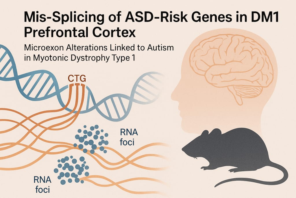
Mis-splicing of ASD-risk genes in DM1 prefrontal cortex
Executive functions are coordinated by the prefrontal cortex, which undergoes transcriptome-wide changes in individuals with ASD25,34. We looked at RNA sequencing (RNA-seq) data from Brodmann area 10 (BA10) of the human DM1 brain to see if the DMPK-CTGexp mutation causes ASD-risk gene RNA mis-splicing in the prefrontal cortex. To reduce age and sex biases, unaffected control samples were matched (Fig. 1a)35,36,37. We calculated the change in "percent spliced in" (PSI) for five AS event types for differential AS analysis (Fig. 1b). 1 percent (1,844) of the 184,000 AS events and 16,000 genes in DM1 met our mis-splicing criteria (PSI > 0.1, FDR 0.05) out of the total pool of 7 percent (1,261) of mis-spliced genes (Extended Data Fig. 1a). All mis-spliced events, including retained intron (RI) events, had a strong positive correlation between CTGexp repeat length and mean PSI values (Fig. 1c and Extended Data Fig. 1b). Consistent with previous research, we observed a negative correlation between the level of intron inclusion and the steady-state level of the host transcript (Extended Data Fig. 1c)38,39, despite the fact that not all RIs introduce a premature termination codon that causes nonsense-mediated decay. We retrieved 38 gene sets and available databases to assess the relevance of DM1 mis-splicing to ASD (Supplementary Table 1), and found that some mis-spliced RI events represented additional RNA species, such as an elevated circular intronic RNA level in DM1 (Extended Data Fig. 1d). After applying more stringent mis-splicing criteria (|PSI| > 0.2), we discovered a significant enrichment of mis-spliced events in 61% of the gene sets (Extended Data Fig. 1e), indicating a consistent trend (Pearson's r = 0.79, P 0.0001). Negative control gene sets (such as genes related to the immune system or metabolism) did not show any enrichment. Notably, the Simons Foundation Autism Research Initiative (SFARI) database and two large Autism Speaks MSSNG-based whole-genome sequencing studies, MSSNG-2017 (ref. 40) and MSSNG-2022 (ref. 40), both showed significant enrichment of ASD-risk genes. 41) (Fig. 1d and Fig. 1e of the Extended Data). SCN2A, ANK2, and SHANK2 were misspliced in DM1 out of 36 overlapping high-confidence ASD-risk genes in both the MSSNG-2017 and MSSNG-2022 studies (Fig. 1e, f, and Extended Data Fig. 1f). In addition, we discovered missplicing in the DMD gene, which is known to be the cause of Duchenne muscular dystrophy and ASD42,43. In addition, we found a strong positive correlation between misspliced events in ASD-risk genes and CTGexp length (Fig. 1g and Extended Data Fig. 1g). These findings, taken as a whole, suggested that the DMPK-CTGexp in the DM1 prefrontal cortex alters the splicing of genes associated with ASD risk.
Mbnl cDKO cortex mimics ASD-risk gene mis-splicing in DM1
The involvement of MBNL splicing factors35 is suggested by the correlation between CTGexp length and the degree of missplicing of ASD-risk genes. We identified the high-affinity MBNL-binding sequences YGCYGCY and YGCY(N)0–5YGCY (where Y denotes pyrimidine) based on our previous research on the MBNL–RNA interaction (14,44,45). In ASD-risk genes in DM1 (FDR = 0.038), a genome-wide distribution analysis revealed a significant enrichment of those binding motifs within 250 bp of mis-spliced skipped/cassette exons (SE) (Fig. 1h). RBPmap46 also predicted that MBNL would bind close to 95% (P = 0.043) of these mis-spliced events (Extended Data Fig. 1h). We used differential AS analysis on RNA-seq data from adult Mbnl1/;Mbnl2c/c;Nestin-Cre+/ conditional double KO mice (hereafter Mbnl cDKO) frontal cortex samples47 to further investigate the role of MBNL in the regulation of ASD-risk genes. Mbnl cDKO mice provide a nervous system-specific model in which Mbnl1 expression is absent in all tissues and Mbnl2 is lost only in the nervous system, including neuronal and glial precursor cells, avoiding the embryonic lethality of constitutive Mbnl1/;Mbnl2/ DKO mice. Missplicing of RNA, altered cortical neuronal and synaptic structures, and widespread brain anatomical changes are all hallmarks of the Mbnl cDKO47,48,49,50. Mis-splicing was found in 13% (1,426) of the genes that were detected, accounting for 5% (1,109) of AS events (Fig. 2a and Extended Data Fig. 2a). In the Mbnl cDKO, mis-splicing was significantly enriched in 61% of gene sets, including ASD-risk gene sets (Fig. 2b and Extended Data Fig. 2b). We detected significant overlaps between SFARI, MSSNG-2017 and MSSNG-2022 mis-spliced ASD-risk genes in human DM1 and mouse Mbnl cDKO cortices (Fig. 2c), which represent distinct systems (such as mutation type). SCN2A was one of 55 ASD-risk genes for which we found a significant overlap (odds ratio (OR) = 1.7, P = 1.6 1012, Fisher’s exact test) (Fig. 2d). All of these findings suggest that ASD-risk gene splicing in the human DM1 cortex is altered by MBNL loss. In DM1 cortical organoids51, the splicing regulator RBFOX1 is downregulated, as are the paralogs of RBFOX in ASD brains27. In human DM1 cortex, our differential gene expression analysis revealed that CTGexp length did not correlate with RBFOX1 RNA level (P = 0.32) (Extended Data Fig. 1i). To explore the impact of RBFOX1 downregulation on mis-splicing of ASD-risk genes in DM1, we retrieved RNA-seq samples from differentiated primary human neural progenitor cells with RBFOX1-knockdown (KD)52. In the RBFOX1 KD, our analysis revealed 235 mis-spliced events (Extended Data Fig. 1j). We found that less than 1% (12) of mis-spliced events in the DM1 overlapped with those observed in RBFOX1 KD cells. The extent of this missplicing was not correlated with CTGexp length (P = 0.62), and ASD-risk genes did not contain any overlapping events. We conducted a genome-wide distribution analysis of the RBFOX1-binding motif (GCAYG) to support this finding. However, we were unable to find any significant enrichment of RBFOX1-binding sequences in mis-spliced ASD-risk genes in DM1 (FDR = 0.38) (Fig. 1h). In addition, RBPmap software predicted no significant RBFOX1 binding in nearby mis-spliced events (P = 0.29). Due to the absence of RBFOX1 involvement, we concluded that missplicing of ASD-risk genes in the DM1 cortex is primarily caused by MBNL loss.
Microexon mis-splicing of ASD-risk genes in DM1 and Mbnl cDKO
In mice, missplicing of neuronal miEs (described here as a 3–33 bp SE), a characteristic of ASD brains, can result in behaviors similar to those of ASD26,30,31. In both the Mbnl cDKO and DM1 cortex, we observed a significant enrichment of miE mis-splicing (Fig. 2e). In the wild-type (WT) mouse frontal cortex, MiEs were found in 4% (n = 1,185) of all SE events, but they were found in 10% (n = 147) of mis-spliced SE events in the Mbnl cDKO, 15% (n = 28) of mis-spliced ASD-risk genes in the SFARI, 23% (n = 6) of MSSNG-2022, and 33% (n = 9) of MSS In a similar vein, miEs accounted for 2% of all detected SE events in the human control prefrontal cortex (n = 3,142), 8% of mis-spliced SE events in DM1, 19% of ASD-risk genes from SFARI (n = 28), 17% of genes from MSSNG-2022 (n = 3) and 47% of genes from MSSNG-2017 (n = 7). In total, we found misspliced miE events in 33 genes, including evolutionarily conserved miEs in high-confidence ASD-risk genes like ANK2, TANC2, and DMD (Fig. 2f,g). These genes are found in both human DM1 and mouse Mbnl cDKO. Comparative in silico modeling of peptides with and without miE-encoded amino acid (aa) sequences was carried out to investigate their potential for protein modulation, as previous research has demonstrated that miEs can locally modify protein structure26. This analysis suggested that some misspliced miEs alter internal protein structures (such as those of ANK2 and NRXN1) or C-terminal protein structures (such as those of DMD and SHANK3) (Fig. 2h and Extended Data Fig. 2c,d). For instance, a protein isoform with a Thr-Ile-Pro (mouse) or Thr-Met-Pro (human) aa sequence is produced when the highly conserved Ank2 miE (12 nt) is included and a proximal alternative 3′ splice site (A3SS) is utilized, whereas miE exclusion promotes the utilization of the distal A3SS (15 nt) and produces a protein isoform with a In myotonic dystrophy, this miE exclusion has been shown to compromise muscle fiber maintenance (Extended Data Fig. 2d)54. For Dmd, a 32-nt miE modifies the structure of the highly conserved dystrophin C terminus that interacts with other proteins. These results show that ASD-risk gene miE splicing is regulated by MBNLs.
ASD-risk gene splicing regulation in cortical development
We looked at the gene expression data for five different mammalian brains at various stages of development55 to see how ASD-risk genes are spliced during development. During the early to middle childhood brain development period, our analysis revealed an increase in MBNL expression that was evolutionary conserved (Fig. 3a). In middle childhood and older brains, MBNL2 expression rises more profoundly and is approximately three times higher than that of MBNL1 (Fig. 3a and Extended Data Fig. 3a), despite the fact that MBNL1 and MBNL2 are expressed at a level that is comparable to that of MBNL1 in the early stages of development and that they both increase simultaneously throughout brain development (Fig. 3a). We looked at RNA-seq data from WT mice at nine developmental time points56 to see if there was a connection between Mbnl1 and Mbnl2 gene expression and MBNL-sensitive splicing transitions in the developing mouse cortex. Mbnl expression levels and mis-splicing levels in ASD-risk genes from the SFARI and both MSSNG studies are strongly correlated, as expected, and this relationship is similar for MBNL-sensitive miEs (Fig. 3b and Extended Data Fig. 3b). 48–56% of mis-spliced AS events in ASD-risk genes were significantly different between the adult cortex and the embryonic cortex, according to differential AS analysis (Extended Data Fig. 3c). Scn2a1 mutually exclusive exons (MXE), Ank2 miE, and Dmd miE splicing transitions, for instance, reached a plateau between two and four weeks of age (Fig. 3c, d, and Extended Data Fig. 3d), which is in line with Mbnl paralogs' developmental expression patterns (Fig. 3a). We analyzed RNA-seq data from primary embryonic cortical neuron samples taken from Mbnl cDKO, constitutive Mbnl1/KO (hereafter Mbnl1 KO), constitutive Mbnl2/KO (hereafter Mbnl2 KO), and WT mice57 to determine whether prenatal MBNL loss influences the splicing of ASD-risk genes. As both Mbnl1 and Mbnl2 are expressed at comparablely low levels in embryonic brains (Fig. 3a and Extended Data Fig. 3a) and they compensate for the loss of each other by regulating the same AS events14, differential splicing analysis did not reveal profound missplicing of ASD-risk genes in single embryonic Mbnl1 KO and Mbnl2 KO. We found significant missplicing of ASD-risk genes, including Dmd miE, in embryonic Mbnl cDKO, as expected (Fig. 3f). This is further supported by the SFARI, MSSNG-2017, and MSSNG-2022 studies' significant enrichment of misspliced events in ASD-risk genes in the DM1 cortical organoids51, including the DMD miE (Fig. 3g,h). In general, these findings suggested that the splicing patterns of numerous ASD-risk genes, including miEs, in the developing brain are governed by MBNL proteins.
Mbnl2 KO causes ASD-risk gene mis-splicing in hippocampus
Mbnl2 is the predominant gene paralog expressed in the adult human and mouse cerebral cortex, hippocampus and cerebellum (Fig. 4a and Extended Data Fig. 4a,b), which are regions known to be involved in ASD58,59. We used RT–PCR splicing analysis of Scn2a MXE, Nrxn1 miE, and Shank3 miE in adult Mbnl2 KO's frontal cortex, hippocampus, and cerebellum to determine whether Mbnl2 loss alters the splicing of ASD-risk genes in multiple brain regions. In two of the three AS events that were tested, missplicing was found to be most prevalent in the hippocampus (Fig. 4b and Extended Data Figs. 4c,d). Consequently, we analyzed differential splicing using RNA-seq data from the hippocampus60 of Mbnl2 KO mice. Scn2a, Ank2, Nrxn1, and Shank3 were among the detected genes that were affected by 4% (n = 912) of AS events (Fig. 4c–e and Extended Data Fig. 4e,f). In the Mbnl2 KO hippocampus, ASD-risk gene lists were significantly enriched among the mis-spliced genes (Fig. 4f and Extended Data Fig. 4g), as was the case with the Mbnl cDKO. We found that 17–25% of overlapping misspliced events are located in ASD-risk genes (Fig. 4g), despite the fact that the overall degree of missplicing varied between the species, models, brain regions, and experiments analyzed (Extended Data Fig. 4h). Scn2a MXE and Ank2 miE were the misspliced events that occurred the most frequently (Fig. 4h). As a result, the AS of ASD-risk genes in multiple adult ASD-relevant brain regions, including the hippocampus, is affected by Mbnl2 loss alone.
MBNLs directly regulate splicing of ASD-relevant microexons
We performed differential AS analysis on RNA-seq data from a mouse brain-derived catecholaminergic (CAD) neuronal cell line with siRNA-mediated Mbnl1;Mbnl2 double KD (hereafter Mbnl DKD)57 to support the observation that MBNLs directly regulate splicing of high-confidence ASD-risk genes. Mis-splicing was found in 5% (n = 2,280) of AS events in 15% (n = 1,536) of detected genes (Fig. 5a and Extended Data Fig. 5a), and mis-splicing was significantly enriched in ASD-risk gene sets (Fig. 5b and Extended Data Fig. 5b). As expected, a genome-wide distribution analysis of high-affinity MBNL-binding sequences identified a significant enrichment of these sequences in mis-spliced ASD-risk genes in all MBNL depletion models (Fig. 5c). We also observed an enrichment of mis-spliced miEs in ASD-risk genes similar to other analyzed mouse model brains, including Ank2 miE (Fig. 5d,e). We selected the highly conserved Ank2 miE, which was consistently mis-spliced in various models, to investigate the mechanism underlying miE splicing because our data indicated that MBNL proteins preferentially regulate miE splicing in ASD-risk genes. We examined MBNL2 crosslinking and immunoprecipitation sequencing (CLIP-seq) samples taken from the hippocampi of adult WT mice60 to determine whether MBNL directly controls Ank2 miE inclusion. Similar to our discovery of an MBNL2-CLIP-seq cluster encompassing a conserved TGCT(N)3TGCT(N)13-18TGCT/C sequence 55 nt downstream of the 5′ splice site in the Ank2/ANK2 intron (Fig. 5f and Extended Data Fig. 5c), previous studies have demonstrated that MBNL-binding to an intron that is downstream of an alternative exon promotes its inclusion60,61. We demonstrated via a filter-binding assay that the recombinant MBNL1 protein does not bind to the RNA with mutated motifs (Kd = 454 1 nM) but does bind to this RNA sequence with high affinity (Kd = 9 4.5 nM). To test whether MBNL can promote alternative exon inclusion by binding to this sequence, we used our previously developed splicing minigene containing MBNL-regulated Atp2a1 exon 22 (E22)44. We substituted the MBNL-binding site in Atp2a1 intron 22 with the normal and mutant mouse Ank2 and human ANK2 conserved intronic sequences (Fig. 5g,h). By PCR with reverse transcription (RT–PCR), we measured the level of E22 inclusion in HeLa cells that had been co-transfected with these Atp2a1 minigenes.
Unlike mutant Ank2/ANK2 and negative control (Δ) minigenes, normal Ank2/ANK2 and positive control (WT) minigenes were sensitive to endogenous MBNLs (Fig. 5h). In addition, normal Ank2/ANK2 contained E22 at significantly higher levels than mutants when Atp2a1 minigenes and MBNL expression vectors were transfected (Fig. 5h and Extended Data Fig. 5e). These results show that MBNLs directly control how Ank2 miE splices an ASD-risk gene.
Mis-splicing of ANK2 microexon in ASD and DM1
We retrieved human adult idiopathic ASD prefrontal cortex (BA9) and matched control samples from the PsychENCODE Consortium (Fig. 6a)24,62 to see if there were any mis-spliced genes or AS events shared by DM1 and ASD. 15 mis-spliced AS events overlapping between DM1 and ASD (OR = 3.5, P = 5.7 105, Fisher’s exact test), including ANK2 miE (Fig. 6c), were identified by our differential AS analysis in 2% (n = 257) of the genes analyzed (Fig. 6b and Extended Data Fig. 6a).
WRITTED BY ME
About the Creator
Mahafuj Alam
🗞️ Mahafuj Alam | News Curator & Independent Media Voice
delivering news quickly, accurately, and bravely, including headline breaking stories and untold local stories. Stay informed. Keep your power.
Keep reading
More stories from Mahafuj Alam and writers in Humans and other communities.
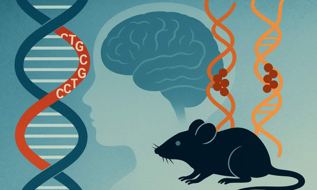
MBNL Sequestration Links Myotonic Dystrophy to Autism via RNA Mis-Splicing
Abstract Autism spectrum disorder has been linked to enrichment of gene-specific tandem repeat expansions across the genome. The CTG tandem repeat expansion in the DMPK gene's 3′ untranslated region, which is known to cause myotonic muscular dystrophy type 1, is one such mutation. Myotonic dystrophy and autism have a clear clinical connection, but the molecular basis for this connection is unknown. During brain development, the RNA splicing patterns of autism-risk genes, particularly a class of autism-relevant microexons, are altered when mutant DMPK RNAs with expanded CUG repeats sequester MBNL splicing factors. We show that both DMPK-CTG expansion and Mbnl null mouse models replicate autism-relevant mis-splicing profiles, as well as altered responses to novelty and social behavioral deficits. Our hypothesis that developmental mis-splicing of autism-risk genes causes myotonic dystrophy-associated autism is supported by these findings.
By Mahafuj Alam10 months ago in Humans
Time to change your life.
Is it even possible for you to change your life? A lot of people, more than there should be, are unhappy with their lives. But what are they doing about it? Usually nothing. People talk about how shitty their lives are and want change, but are they not taking the actions to change?
By Jen Phillipsa day ago in Humans
Heightened Intuition and How It Shows in Decision Making.
Intuition is a form of inner knowing that guides decisions without relying solely on logical reasoning. People with heightened intuition often detect patterns, anticipate outcomes, and make choices that align with both immediate circumstances and long-term goals. This ability is not magical; it reflects the brain’s capacity to process complex information quickly, drawing from experience, memory, and subconscious cues. Understanding how intuition manifests in decision making allows individuals to leverage it effectively in both personal and professional life.
By Wilson Igbasiabout 6 hours ago in Humans
One Unchecked Box
"Republished" because it was the only way to add the embed for the newly recorded audio version of this story due to the Top Story badge. Plus it serves as a nice, informal announcement of the podcast's revival for another season (go subscribe!):
By Stephen A. Roddewig5 days ago in Fiction


Comments
There are no comments for this story
Be the first to respond and start the conversation.