
Top 10 Signs of Inflammation in Brain MRI Scans
Top 10 Indicators of Inflammation in Brain MRI Scans
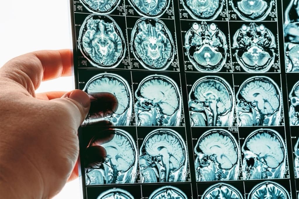
Inflammation in the brain is a serious condition that can lead to significant health problems. An MRI scan serves as a vital tool in diagnosing this inflammation. MRI technology captures detailed images of the brain, revealing subtle changes that may indicate underlying issues. This article explores the top 10 signs of inflammation in brain MRI scans, helping you understand what to look for and why these signs matter.
Identifying Lesions
Lesions often appear as dark or light spots on MRI scans, signifying potential inflammation. These lesions may develop in various brain regions, indicating areas of damage or abnormality. Clinicians assess these lesions' size, shape, and location to determine their significance. Furthermore, they can differentiate between active inflammation and previous scarring. Recognizing these lesions early allows for timely intervention and management. Patients should be aware that not all lesions indicate inflammation; thus, further evaluation is often necessary to understand their nature fully.
Abnormalities in White Matter
White matter abnormalities are common signs of inflammation visible in brain MRI scans. These changes often manifest as hyperintense regions, suggesting areas where the myelin sheath—the protective covering around nerve fibers—has deteriorated. Clinicians frequently associate white matter lesions with multiple sclerosis and other inflammatory diseases. Additionally, these abnormalities can help gauge the disease's progression and response to treatment. Identifying these changes early in patients provides essential insights into their neurological health and aids in developing effective treatment plans.
Swelling in Specific Brain Regions
Swelling in the brain, known as edema, frequently appears on MRI scans as a clear indicator of inflammation. Clinicians pay close attention to regions such as the cortex and subcortical areas where swelling can occur. Furthermore, localized swelling often correlates with specific conditions, such as infections or autoimmune disorders. It signifies the body's response to injury or illness. Detecting swelling allows for timely diagnosis and intervention, crucial in preventing further complications. Additionally, monitoring the progression or resolution of swelling informs treatment effectiveness. Many individuals find that books on self care help them manage stress related to these health concerns.
Increased Signal Intensity
Increased signal intensity on MRI scans can indicate inflammation in the brain. This heightened signal often reflects the presence of fluid or other substances related to inflammatory processes. Clinicians evaluate these changes carefully, as they can help identify active inflammation versus chronic changes. Furthermore, increased signal intensity can also highlight areas of demyelination or vascular issues, adding complexity to the diagnosis. Understanding the implications of increased signal intensity assists healthcare providers in forming accurate assessments and treatment plans for patients.
Contrast Enhancement
Contrast enhancement plays a crucial role in revealing inflammation during MRI scans. When a contrast agent is injected, it highlights areas of abnormal vascular permeability associated with inflammation. Clinicians look for enhanced areas, as they often indicate active inflammation, tumors, or infections. Moreover, the degree of enhancement can help determine the severity and extent of the condition. Additionally, contrast-enhanced MRI provides valuable information about the blood-brain barrier's integrity, offering insights into the underlying mechanisms of disease. Therefore, it becomes an essential part of the diagnostic process.
Changes in Cerebrospinal Fluid
Cerebrospinal fluid (CSF) changes observed on MRI scans can indicate inflammation in the brain. Clinicians assess the CSF for abnormalities, including increased protein levels or the presence of inflammatory cells. Additionally, changes in the appearance of the ventricles—fluid-filled spaces in the brain—can signal potential issues. For instance, enlargement of the ventricles may suggest an accumulation of CSF due to inflammation. Identifying these changes is crucial for diagnosing conditions like meningitis or encephalitis. Monitoring CSF changes informs treatment decisions and helps evaluate the patient's response to therapy.
Vascular Changes
Vascular changes visible on MRI scans can reveal important information about brain inflammation. These changes may include blood flow or vessel structure alterations, suggesting underlying inflammatory processes. For example, abnormalities in the veins or arteries can indicate conditions like vasculitis, where blood vessels become inflamed. Furthermore, identifying vascular changes helps clinicians understand the extent of inflammation and its potential impact on brain function. By recognizing these changes, healthcare providers can develop targeted treatment strategies to address the inflammation and its effects on the vascular system.
Focal or Diffuse Patterns
MRI scans can display focal or diffuse patterns of inflammation, each with distinct implications. Focal patterns indicate localized inflammation, often associated with specific conditions such as abscesses or tumors. In contrast, diffuse patterns suggest widespread inflammation, commonly seen in autoimmune disorders. Clinicians carefully analyze these patterns to inform their diagnostic and treatment approaches. Additionally, recognizing the pattern of inflammation can aid in predicting disease progression and response to therapy. Thus, understanding the significance of focal versus diffuse patterns is essential in managing brain inflammation effectively.
Correlating Clinical Symptoms with MRI Findings
Correlating clinical symptoms with MRI findings is crucial for accurate diagnosis. Patients often present with neurological symptoms, such as headaches, cognitive changes, or motor deficits. Clinicians compare these symptoms with MRI results to comprehensively understand the patient's condition. Furthermore, this correlation helps distinguish between the causes of inflammation and guides treatment decisions. For instance, specific MRI signs combined with certain symptoms can indicate conditions like multiple sclerosis or encephalitis. Therefore, a thorough evaluation of both clinical and imaging data is essential.
When to Seek Further Evaluation
Knowing when to seek further evaluation for inflammation in brain MRI is essential for timely diagnosis and treatment. Patients with abnormal MRI findings or persistent neurological symptoms should consult their healthcare providers for a comprehensive assessment. Additionally, follow-up imaging studies may be necessary to monitor changes over time or evaluate the effectiveness of treatment. Clinicians may also recommend additional tests, such as lumbar punctures or blood tests, to gain further insights into the underlying causes of inflammation. Early intervention can significantly impact patient outcomes and overall quality of life.
Conclusion
Understanding the signs of inflammation in brain MRI scans is vital for early diagnosis and effective treatment. Each sign provides crucial insights into the patient's condition, from identifying lesions to recognizing vascular changes. Additionally, correlating clinical symptoms with MRI findings enhances diagnostic accuracy. By being aware of these signs, patients and healthcare providers can work together to manage inflammation effectively. Ultimately, timely intervention can lead to better outcomes and improved quality of life for individuals affected by brain inflammation.
About the Creator
Keep reading
More stories from roberto andreas and writers in Writers and other communities.
Delicious Recipes & Meal Ideas from Author of Gluten Free Burnout
Living a gluten-free lifestyle can be both rewarding and challenging. The author of Gluten Free Burnout has created a wealth of delicious recipes that make this journey enjoyable and satisfying. In this blog, we’ll look at some mouthwatering meal ideas that not only showcase the benefits of a gluten free lifestyle but also inspire creativity in the kitchen. Whether you're new to gluten-free eating or a seasoned veteran, these recipes will make you excited about your meals!
By roberto andreasabout a year ago in Feast


Comments
There are no comments for this story
Be the first to respond and start the conversation.