
The organisation of behaviour is revealed by brain activity.
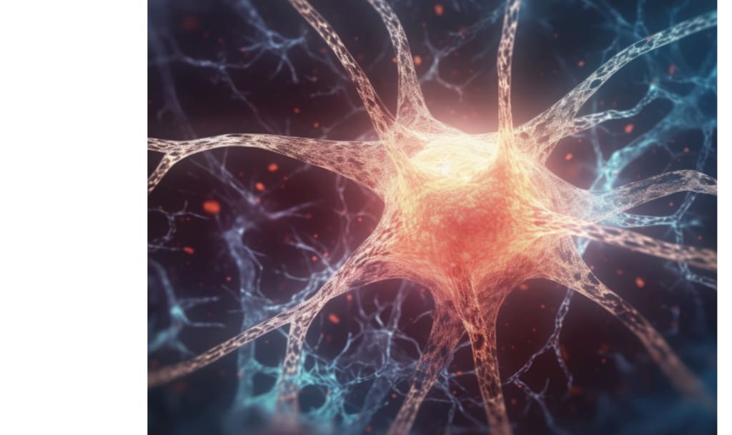
Brain mapping for behaviour

Based on how brain tissue appears under a microscope, scientists have been classifying the brain into regions for more than a century.
The maps have influenced how scientists investigate cognition, judgement, and self-control, particularly in the prefrontal cortex of the brain. According to a recent study, those maps might not capture the whole picture.
The way neurons fire together over time appears to organise the brain's control circuits rather than aligning with obvious tissue boundaries. To put it another way, the behaviour of cells is more important than their location.
Researchers discovered that brain processes and behaviours like planning and decision-making cluster according to similar activity patterns by monitoring the activity of tens of thousands of neurones in awake mice.
The research, headed by Marie Carlén at Karolinska Institutet, demonstrates that neural activity, not conventional anatomical labelling, organises the prefrontal cortex, changing scientists' perceptions of the brain's most crucial regulatory region.
Brain mapping for behaviour
Traditional atlases began with thin brain tissue slices that were dyed to make the various layers stand out in the light. The cell arrangement visible in dyed slices was referred to by scientists as cytoarchitecture, which they used to identify different parts of the brain.
According to a 2018 assessment, Brodmann-style maps have influenced contemporary research for over a century. Even when behaviour expanded across bigger networks, it became simple to consider each region as though it performed a distinct task since those borders appear sharp.
Live recording of brain activity
Individual neurones, which communicate by transmitting brief electrical signals to neighbouring cells, were recorded by Carlén's team using small electrodes. Because it resembles how local circuits often function, they concentrate on spontaneous activity, or firing that persists without a clear trigger.
Some neurones fired regularly and slowly across 3-second periods, whereas others fired rapidly and in spurts. The researchers were able to draw borders according to function by mapping those patterns onto the locations of each neurone.
The order of the brain that directs behaviour
According to a well-known notion, the prefrontal cortex is close to the top of the brain's information organisation hierarchy and is associated with goal-directed control.
Subsequent research in 2019 connected the way the cortex and thalamus connect and transmit signals to cortical hierarchy, or ranked levels of information flow.
The findings demonstrated that activity patterns tracked that ranking more accurately than they did the typical tissue borders. According to this link, even when adjacent tissue seems comparable under a microscope, wiring may influence what neurones do.
Some neurones fire steadily.
High in the prefrontal hierarchy, a distinct signature was evident: a large number of neurones fired steadily and at a low rate. Recurrent connection, where signals loop across local circuits to aid in information integration over time, is probably reflected in this regular activity.
Slow and steady firing also identified high-ranking areas within the internal organisation of the mouse prefrontal cortex. Disruptions to that rhythm may impair planning and reasoning before issues become apparent if these neurones aid in maintaining information.
Quick dismissal while considering decisions
Concurrently, those identical high-ranking areas' decision-related neurones exhibited drastically different behaviours. They fired quickly instead than gradually, a pattern that is ideal for encoding decisions via varying spike counts in milliseconds.
According to Carlén, "some neurones appear to specialise in integrating information streams, while others have high spontaneous activity that supports quick and flexible encoding of information, such as information needed to make a specific decision."
The findings collectively suggest local specialisation in the prefrontal cortex, where distinct firing patterns serve various cognitive functions while individual neurones maintain flexibility in response to changing task demands.
Behaviour and brain maps don't match
Experiments frequently focus on a single label, and traditional labels treat the prefrontal cortex as a collection of identified sections. The new maps separated space according to firing patterns, so a module may split or cross two designated areas.
Borders appeared less trustworthy in their recordings because tone responses and choice signals landed in patchy areas. New methods for reporting locations will be required for future work, otherwise minor disputes across labs will continue to accumulate.
Neural detail-capturing tools
Hardware that samples many sites simultaneously without inhibiting behaviour was needed to record from tens of thousands of cells. Neuropixels, silicon probes with hundreds of recording locations that record neural spikes over extended regions of brain tissue, were a significant advancement.
Following the separation of mixed electrical traces into individual neurones by computers, mapping software assigned those neurones to common coordinates. Labs are able to compare activity maps between different brain areas, animals, and even disease models since that pipeline scales.
Mental disease and brain control
Planning, impulse control, and mood regulation—functions historically associated with the prefrontal cortex—are impaired in many mental diseases.
These illnesses share prefrontal deficits, according to a large human analysis, and the intensity of symptoms closely correlates with reduced activity in circuits related to cognitive control.
Carlén noted, "It is surprising how little is known about how this region actually works, considering that deviations in prefrontal cortex function have been linked to virtually all psychiatric disorders."
When considered collectively, the results indicate that neuronal activity patterns establish useful functional boundaries in the prefrontal cortex more precisely than tissue appearance alone.
Researchers may now examine how certain prefrontal modules alter in response to stress, medication, or illness while maintaining anatomical anchors thanks to these activity-based maps.
However, the scientists warn that human symptoms cannot be predicted by mouse-based insights alone, thus careful translation and validation will continue to be crucial.
About the Creator
Keep reading
More stories from Francis Dami and writers in Psyche and other communities.
🅼🅸🅳🅽🅸🅶🅷🆃 🆂🅽🅰🅲🅺🆂
"It's 10 in Tuscon! We all know what that means... It's Time for Midnight Snacks with your man, Gerald Gee! Ready to spend the night together? Me too! I'm full of snacks and can't wait to regurgitate them all back into your hungry ears. Crack a brew! Pop some corn! Anything to get ready for one hell of a show where the talk maybe cheap but the words cut deep...
By Lamar Wiggins5 days ago in Fiction


Comments
There are no comments for this story
Be the first to respond and start the conversation.