
The Role of Vitrectomy in Treating Complex Eye Conditions
Vitrectomy for Complex Eye Conditions


Our eyes provide us with a window to the world, but complex conditions can sometimes cloud that view. When issues arise in the delicate, jelly-like substance filling the eyeball, known as the vitreous humor, a specialized procedure is often required. An eye surgery vitrectomy is a sophisticated technique used by retinal specialists to treat a wide range of problems affecting the back of the eye, helping to restore, protect, and improve vision for countless patients. This procedure involves the careful removal of the vitreous humor, allowing surgeons to access the retina and make necessary repairs.
Understanding this procedure is the first step toward appreciating its impact on modern eye care. It’s more than just a surgery; it’s a pathway to clearer sight for those facing significant visual challenges. We will explore what a vitrectomy entails, the conditions it addresses, and what patients can expect during the recovery process.
What is a Vitrectomy?
A vitrectomy is a type of microsurgery performed by a retina specialist. The primary goal is to remove some or all of the vitreous humor from the eye. This clear, gel-like substance fills the large space in the middle of the eyeball, behind the lens. It helps the eye maintain its round shape and contains fine fibers that are attached to the retina, the light-sensitive tissue at the back of the eye.
Over time, or due to disease or injury, the vitreous can change. It might shrink, pull on the retina, become cloudy with blood or debris, or harbor scar tissue. Removing it gives the surgeon unobstructed access to the retina and macula, allowing for precise treatment of various disorders.
The Surgical Process: A Step-by-Step Overview
A vitrectomy is typically performed as an outpatient procedure under local or general anesthesia. The surgeon makes several tiny incisions in the sclera (the white part of the eye) to insert microscopic instruments.
- Step 1: Infusion: A port introduces a sterile saline solution to maintain the eye's pressure and shape once the vitreous is removed.
- Step 2: Illumination: A fiber-optic light pipe is inserted to illuminate the inside of the eye, giving the surgeon a clear view.
- Step 3: Vitrector: A tiny cutting instrument, the vitrector, is used to carefully cut and remove the vitreous gel.
- Step 4: Repair: With the vitreous gone, the surgeon can perform necessary repairs, such as fixing a retinal tear or removing scar tissue.
- Step 5: Replacement: The eye is refilled with a substitute for the vitreous. This can be a saline solution, a gas bubble, or silicone oil, depending on the condition being treated.
Conditions Treated with Vitrectomy
This procedure is a versatile tool for addressing numerous complex issues. The use of vitrectomy for eye conditions has become a standard of care when other treatments are not sufficient.
Retinal Detachment
A retinal detachment is a serious emergency where the retina pulls away from its normal position. If not treated quickly, it can cause permanent vision loss. A vitrectomy serves as an effective retinal detachment treatment by removing the vitreous that is pulling on the retina. The surgeon can then use a laser or cryotherapy (a freezing technique) to seal the retinal tears and reattach the retina, often with the help of a gas bubble to hold it in place.
Diabetic Retinopathy
For individuals with advanced diabetes, abnormal blood vessels can grow on the retina. These vessels can leak blood into the vitreous (vitreous hemorrhage) or form scar tissue that can detach the retina. A diabetic retinopathy vitrectomy is performed to clear the blood-filled vitreous and remove the scar tissue, helping to prevent further vision loss and stabilize the eye.
Did You Know? According to the National Eye Institute, diabetic retinopathy is the leading cause of blindness in American adults. Early detection and advanced treatments like vitrectomy are crucial in managing the disease.
Macular Holes and Puckers
The macula is the central part of the retina responsible for sharp, detailed vision. A macular hole is a small break in the macula, while a macular pucker is scar tissue that has formed over it. Both can cause blurred and distorted central vision. During a vitrectomy, the surgeon can remove the vitreous and the membrane causing the pucker or hole, often improving vision significantly.
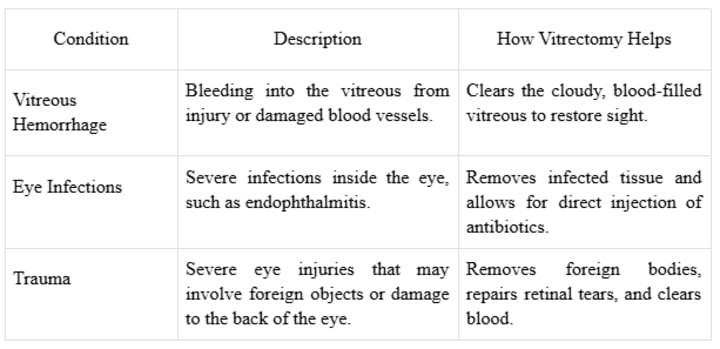
Other Applications
A vitrectomy is also a valuable complex eye surgery treatment for several other issues:
Recovery and Expectations After Surgery
Recovery from a vitrectomy varies depending on the specific reason for the surgery and the substance used to replace the vitreous.
- Saline Solution: If the eye is filled with saline, the body will naturally replace it with its own fluid (aqueous humor) over time. Vision typically improves gradually over several weeks.
- Gas Bubble: If a gas bubble is used, you may be required to maintain a specific head position (often face-down) for several days or weeks. This allows the bubble to press against the retina and hold it in place while it heals. Vision will be very poor until the bubble is naturally absorbed by the body. Air travel is strictly forbidden until the gas bubble is gone.
- Silicone Oil: Silicone oil provides longer-term support for the retina. It does not get absorbed and usually requires a second, minor surgery for its removal months later.
Pro Tip: Prepare a "recovery station" before your surgery. If face-down positioning is required, rent or purchase specialized equipment like a vitrectomy chair, mirror, or pillows to make the period more comfortable.
Post-surgery, patients will use prescribed eye drops to prevent infection and reduce inflammation. It's normal to experience some discomfort, redness, and blurry vision. Following all post-operative instructions from your surgeon is essential for a successful outcome.
Final Words
A vitrectomy is a powerful procedure for treating complex eye diseases that affect the retina and vitreous.
- It's a versatile surgery: It can address a wide range of issues, from diabetic retinopathy and retinal detachments to macular holes and eye trauma.
- It provides access: By removing the vitreous, surgeons can directly repair damage to the retina.
- Recovery is crucial: The outcome depends heavily on the specific procedure performed and the patient's adherence to post-operative care, especially head positioning if a gas bubble is used.
- It restores vision: For many, a vitrectomy can halt progressive vision loss and, in many cases, restore functional sight.
A vitrectomy represents a significant advancement in ophthalmology, offering hope and healing to individuals facing serious threats to their vision. By understanding its role, patients can feel more empowered and prepared when discussing treatment options with their eye care specialist.
FAQs
1. Is a vitrectomy painful?
The surgery itself is not painful, as it is performed under local or general anesthesia. Post-operative discomfort is common but can usually be managed with prescribed pain medication and eye drops.
2. How long does it take to recover from a vitrectomy?
Full recovery can take several weeks to months. The initial healing phase is typically a few weeks, but visual recovery depends on the underlying condition and the substance (saline, gas, or oil) used to fill the eye.
3. Will my vision be perfect after a vitrectomy?
The goal of a vitrectomy is to stabilize the eye and prevent further vision loss, with the hope of improvement. The final visual outcome depends on the extent of the damage to the retina before the surgery. While many patients experience significant visual improvement, some may still have limitations.
About the Creator
Franklin Norton
Franklin Norton is a dedicated health writer specializing in eye care. With a passion for vision health, he educates readers on eye conditions, treatments, and preventive care for optimal eye wellness.
Keep reading
More stories from Franklin Norton and writers in Education and other communities.
Dry Macular Degeneration Treatment: Exploring FDA Innovations
Living with dry macular degeneration can feel like watching the world slowly dim. Central vision, crucial for reading, driving, and recognizing faces, gradually blurs. For many years, treatment options were limited, focusing on slowing the disease's progression rather than halting or reversing its effects. However, the landscape is changing. Recent breakthroughs have led to new, FDA-approved therapies, offering renewed hope to millions. This article will explore these innovative treatments, explaining how they work and what they mean for people managing this condition.
By Franklin Norton4 months ago in Education
Traphall Romance
Traphall Romance: How a Global Love Sound Was Born, Who’s Leading It, and Where It’s Headed Genres don’t usually start in boardrooms. They start in bedrooms, cars, late-night studio sessions, and cities where cultures overlap. Traphall Romance is no exception. It’s not a sound that was formally announced — it’s one that quietly emerged from emotion, from artists who wanted their music to hit hard and feel deep.
By K.y.e Dynasty Recordsabout 20 hours ago in Education
How Top-Ranked Teams Build a Culture of Excellence and Consistent High Performance
In today’s competitive business environment, success is rarely the result of individual talent alone. Strong, aligned, and resilient teams power organizations that consistently lead their industries. Lists highlighting outstanding teams, such as those curated by Forbes, have drawn attention to a critical truth: exceptional performance is built intentionally. These teams succeed because of how they collaborate, lead, and adapt over time. Understanding what sets these high-performing teams apart provides a roadmap for organizations seeking sustainable excellence.
By Daniel Ladner2 days ago in Education


Comments
There are no comments for this story
Be the first to respond and start the conversation.