
Exploring the Endoplasmic Reticulum: The Cell’s Internal Highway System
Structure and Significance of the Endoplasmic Reticulum in Cellular Function


Imagine walking into a massive factory with conveyor belts running in every direction—moving, folding, and transporting essential materials. Inside every cell, a similar structure exists: the Endoplasmic Reticulum (ER). It's a vital, membrane-bound organelle that plays a central role in the cell’s internal transport system.
The term Endoplasmic Reticulum itself reveals a lot about its nature:
"Endo" means inside,
"Plasmic" refers to the cytoplasm, and
"Reticulum" means network.
So, the ER is essentially a network inside the cytoplasm—a vast system of folded membranes that helps the cell stay organized and productive.
Structure at a Glance:
The Endoplasmic Reticulum is not just a random jumble. It is a highly structured organelle made of membranes composed of proteins and lipids. These membranes form folded structures—increasing surface area for efficient functioning.
It comes in three types of structures:
Cisternae – flat, sac-like structures
Vesicles – small, round, bubble-like parts
Tubules – tube-shaped channels
Cisternae – The Flat Sacs of the ER:
This slide focuses on the Cisternae, the most recognizable part of the ER. These are flattened sacs that are often stacked upon each other. Inside them is a fluid-filled space called the lumen, where chemical processes occur.
The Ribosomes attached to the surface give it a “rough” appearance in the Rough ER, while Smooth ER lacks ribosomes. Special proteins like Ribophorin help in the attachment of ribosomes and processing of proteins.
The cisternae are interconnected, forming a continuous system that runs throughout the cell—making it a true network of communication and processing.
Dimensions Matter:
The ER spans a diameter of about 50-60 micrometers, allowing it to cover a significant portion of the cytoplasm.
The Endoplasmic Matrix, a fluid within the ER, supports all internal transport and reactions.
This foundational understanding sets the stage for exploring how the ER operates in detail—how it helps produce proteins, lipids, and transports vital molecules. Stay tuned for the next slides where we’ll dig into the types of ER, their functions, and biological importance.

Vesicles and Tubules: The Dynamic Arms of the Endoplasmic Reticulum
As we dive deeper into the ''architecture of the Endoplasmic Reticulum (ER)'', we encounter two more essential components beyond the flat cisternae: Vesicles and Tubules.
These structures, though smaller and more flexible, perform massive roles in ensuring that proteins, lipids, and other molecules are safely processed, packaged, and sent to their destinations inside or outside the cell.
Vesicles – The Cellular Delivery Bubbles:
Think of Vesicles as tiny, membrane-bound sacs that form near the cisternae. Their main job? ''Transport''.
Just like sealed packages leaving a factory, vesicles carry substances produced or processed by the ER to other organelles, or sometimes to the plasma membrane for secretion.
They are extremely vital for:
Intracellular transport
Temporary storage
Protein modification and sorting
Their formation ensures that no precious cargo is lost during the transfer process within the bustling environment of the cytoplasm.
Tubules – The Flexible Branches of the ER:
Next up, we have Tubules—branch-like structures that stretch across the cell, closer to the plasma membrane. Unlike the flat cisternae, tubules have a more cylindrical and narrow shape, with diameters ranging from 50 to 500 micrometers.
Their main characteristics:
Do not have ribosomes
Are more prominent in the Smooth ER
Facilitate lipid synthesis and detoxification
Help connect different regions of the ER and extend toward the plasma membrane
Because they lack ribosomes, tubules are associated more with smooth ER functions, like making hormones, breaking down toxins, and regulating calcium levels.
Both vesicles and tubules may appear small, but their roles in cellular homeostasis are huge. Together with cisternae, they create a versatile and efficient network that adapts to the needs of the cell in real time.
TYPES OF ER (Endoplasmic Reticulum)
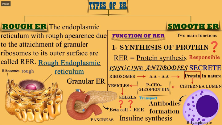
1. Rough ER (RER)
Definition: The Rough Endoplasmic Reticulum is called rough because of the presence of ribosomes on its surface, giving it a granular appearance.
Alternate Name: Also referred to as Granular ER.
Function:
Protein Synthesis: It synthesizes proteins, especially those that are secreted out of the cell like insulin and antibodies.
Ribosomes (attached to RER) create polypeptides by linking amino acids (A.A).
These proteins are synthesized into the cisternal lumen of the RER.
Proteins may be modified into glycoproteins (proteins + carbohydrates).
Vesicles transport proteins to the Golgi apparatus for further processing.
Specific Examples Given: Insulin synthesis by beta cells of the pancreas (highlighted on the slide).
Antibody productionby B lymphocytes (immune cells).
Key Takeaway: The Rough ER plays a critical role in synthesizing proteins, particularly secretory proteins like insulin and antibodies. These proteins are processed through the cisternae, packaged into vesicles, and sent to the Golgi apparatus for further modification and distribution.
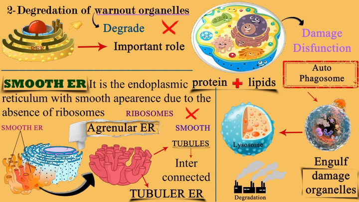
Degradation of Worn-Out Organelles:
While the RER does not directly degrade worn-out organelles, it plays a critical indirect role in the cellular degradation system, especially autophagy. Here's how:
Step-by-Step Explanation:
1. Protein Synthesis for Lysosomes
The RER synthesizes proteins, especially enzymes, that are essential for lysosome formation. These proteins include hydrolytic enzymes that can break down cellular components.
2. Transport via Golgi Apparatus
These enzymes are then transported from the RER to the Golgi apparatus, where they are processed and packaged into lysosomes.
3. Lysosome Formation
The lysosomes—which contain these enzymes—are the organelles directly responsible for degrading worn-out or damaged organelles, misfolded proteins, and other cellular debris.
4. Autophagy Process
When an organelle becomes dysfunctional, it is engulfed by a membrane, forming an autophagosome.
This autophagosome then fuses with a lysosome, and the contents are degraded.
Summary:
The RER supports degradation of worn-out organelles by producing the hydrolytic enzymes required by lysosomes.
These lysosomes then carry out autophagy—the actual process of degrading old or damaged organelles.
Think of the Rough ER as the factory that makes the tools (enzymes), while the lysosome is the janitor that uses those tools to clean up the cell.
2. Smooth Endoplasmic Reticulum (SER)
Definition: Smooth ER is a type of endoplasmic reticulum that lacks ribosomes, giving it a smooth appearance.
Alternate name: Agranular ER.
Structural Features:
Composed of interconnected tubules → labeled as Tubular ER.
Lacks ribosomes, which distinguishes it from RER.
Keywords Used:
SMOOTH, TUBULES, Inter connected, TUBULAR ER
The Smooth ER is primarily involved in lipid synthesis and detoxification, with a distinct lack of ribosomes.
The cell’s waste disposal system involves autophagy where autophagosomes engulf damaged organelles and fuse with lysosomes for degradation, keeping the cell healthy and functioning.
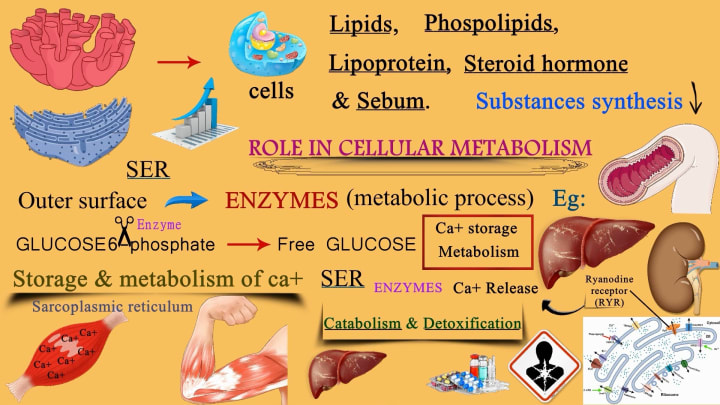
Functions of SER:
1. The SER is responsible for the synthesis of
Lipids
Phospholipids
Lipoproteins
Steroid hormones
Sebum (an oily secretion from sebaceous glands)
These are essential for:
Building cell membranes
Hormone production
Protecting and lubricating tissues
2. Role in Cellular Metabolism
The central message: SER plays a vital role in cellular metabolism through enzyme action and biochemical pathways.
Highlighted areas include:
Enzymes (metabolic process):
Located on the outer surface of the SER.
These enzymes help in various metabolic processes, such as glucose metabolism.
Example shown:
Glucose-6-phosphate is broken down by an enzyme into free glucose, which can then be used by cells for energy.
3. Calcium (Ca²⁺) Storage and Metabolism
SER regulates calcium ion (Ca²⁺) storage, especially in muscle cells, where it’s called the sarcoplasmic reticulum.
Ca²⁺ is stored inside the SER.
When needed (e.g., for muscle contraction), Ca²⁺ is released via Ryanodine Receptors (RYR).
This process is crucial for muscle function and signaling.
Muscle arm + sarcoplasmic reticulum = calcium storage/release for muscle activity
Liver and kidney also shown as examples where calcium metabolism occurs
4. Catabolism & Detoxification
SER is heavily involved in detoxifying harmful substances (e.g., drugs, poisons).
Catabolism refers to breaking down molecules to extract energy.
Enzymes in the SER:
Modify toxic compounds (like alcohol or drugs) into safer substances
Help maintain liver and kidney function.
About the Creator
EasyMedEdHub
Welcome to EasyMedEdHub! 🎓
EasyMedEdHub simplifies complex science and medical concepts with clear, engaging content. Follow for tips, insights, and resources to help you succeed academically in biology and beyond!
Keep reading
More stories from EasyMedEdHub and writers in Education and other communities.
Cytosol: The Invisible Engine Powering Every Cell
slide 1 What is Cytoplasm? The Gel That Keeps Your Cells Alive When we think about cells, we often picture the nucleus — the control center. But what about the space between the nucleus and the outer cell membrane? That jelly-like substance that holds everything together and keeps your cells functioning smoothly? That’s called cytoplasm, and it's more important than you might think.
By EasyMedEdHub10 months ago in Education
The Red Renaissance Uncorking the Future of Russian Wine in 2026
For decades, the global wine map was a predictable atlas of French Chateaus, Italian vineyards, and New World giants. However, as we move through 2026, a new and unexpected frontier has firmly established its presence: Russia. The Russian wine industry is currently undergoing a "Grand Renaissance," fueled by a combination of aggressive state support, massive private investment, and a geopolitical shift that has turned the domestic market inward. What was once seen as a secondary player, primarily known for mass-produced sparkling wines, has transformed into a sophisticated producer of premium, terroir-driven still wines that are winning over a new generation of health-conscious and patriotic consumers.
By Neeraj kumar4 days ago in Education
Cabin Fever Because of Snow, Sleet, and Freezing Rain That Turned to Icy Roads
What Is Cabin Fever? The short answer is that cabin fever is restlessness from being in a confined area. Cabin fever is the distressing irritability or restlessness experienced when a person or group is stuck at an isolated location or in confined quarters for an extended time. Research shows that prolonged cold, gray skies, and being stuck indoors can trigger mood shifts similar to “winter blues.”
By Margaret Minnicks2 days ago in Humans


Comments
There are no comments for this story
Be the first to respond and start the conversation.