
Unbeknownst Anomalies: 10 Unusual Traits You Could Have
Unaware of the Anomalies!
Have you ever wondered about the hidden anomalies that may exist within your own body? The human body is a fascinating entity, filled with complexities and mysteries waiting to be unraveled. While most of us are familiar with common physical characteristics, there are some extraordinary traits that remain unnoticed until discovered. In this article, we will explore ten fascinating anomalies that you might possess without even realizing it
Situs Inversus: Could Your Internal Organs be Reversed?
The human body is a complex and intricate system, with each organ playing a specific role in maintaining our health and well-being. While most individuals have their internal organs arranged in a predictable pattern, there are rare cases where an extraordinary phenomenon called situs inversus occurs. Situs inversus is a condition in which the internal organs are reversed or mirrored from their normal positions. In this article, we will explore situs inversus, its causes, implications, and how it affects those who have this unique condition.
Understanding Situs Inversus:
Situs inversus, also known as "mirror-image reversal," occurs during embryonic development when the internal organs, such as the heart, liver, stomach, and intestines, form in a reversed pattern. In individuals with situs inversus, the heart, usually located on the left side of the chest, is found on the right side, while the liver, typically situated on the right side, is found on the left side. This reversal affects the entire arrangement of the internal organs, creating a mirror-image configuration.
Causes and Types of Situs Inversus:
The exact cause of situs inversus is not yet fully understood. However, researchers believe that genetic factors play a significant role in its development. Situs inversus can be classified into two types: situs inversus totalis and situs inversus partialis. Situs inversus totalis refers to the complete reversal of all internal organs, while situs inversus partialis involves the reversal of only a specific subset of organs.
Implications and Medical Challenges:
While situs inversus itself is not a life-threatening condition, it can present some challenges in medical situations. Healthcare professionals need to be aware of this condition to avoid potential diagnostic errors or complications during surgeries. For example, when performing an appendectomy on a patient with situs inversus, surgeons must adapt their surgical approach due to the reversed position of the appendix.
Discovering Situs Inversus:
In many cases, situs inversus goes undiagnosed until a medical examination or imaging scan reveals the unusual arrangement of internal organs. Some individuals may live their entire lives without realizing they have this condition unless it becomes incidentally discovered during routine medical procedures or tests.
Living with Situs Inversus:
Fortunately, situs inversus does not typically cause any health issues or impair organ function. People with situs inversus can lead normal, healthy lives, just like individuals without this condition. However, it is essential for individuals with situs inversus to inform their healthcare providers about their condition to ensure accurate diagnoses and appropriate medical treatments.
Situs Inversus as a Genetic Marker:
Interestingly, situs inversus can be an indicator of an underlying genetic condition called primary ciliary dyskinesia (PCD). PCD is a rare genetic disorder that affects the movement of cilia, tiny hair-like structures that line the respiratory tract, fallopian tubes, and other parts of the body. Situs inversus is found in approximately 50% of individuals with PCD, highlighting the genetic link between these two conditions.
Conclusion:
Situs inversus is a fascinating condition that challenges our understanding of human anatomy. While it may seem peculiar to have reversed internal organs, individuals with situs inversus can live normal lives without significant health consequences. This condition serves as a reminder of the incredible diversity and complexity of the human body, offering valuable insights into embryonic development and genetic factors that shape our physical characteristics.
Hammer Toe Deformity: When Testicles Swing Excessively!
The human body is a fascinating and complex system, with various parts and organs serving different functions. However, sometimes unusual conditions or deformities can occur, leading to intriguing and sometimes uncomfortable situations. One such condition is hammer toe deformity, which affects the testicles, causing them to swing excessively. In this article, we will explore the causes, symptoms, and potential treatment options for hammer toe deformity.
Understanding Hammer Toe Deformity:
Hammer toe deformity, also known as testicular hypermobility or testicular torsion, is a condition in which the testicles swing more than usual. Normally, the testicles are held in place by a network of tissues and muscles within the scrotum. However, in cases of hammer toe deformity, this support system weakens or becomes lax, allowing the testicles to move excessively.
Causes of Hammer Toe Deformity:
Hammer toe deformity can have several potential causes. It can be congenital, meaning it is present from birth and results from abnormal development of the scrotum and its supporting structures. In other cases, it may be acquired due to factors such as trauma or injury to the scrotum, hormonal imbalances, or weakening of the supporting tissues due to age or certain medical conditions.
Symptoms and Effects:
The main symptom of hammer toe deformity is the excessive swinging or movement of the testicles. This can cause discomfort or pain, especially during physical activities or when wearing loose-fitting underwear. Some individuals may also experience embarrassment or self-consciousness due to the visible movement of their testicles.
Treatment Options:
The treatment for hammer toe deformity depends on the severity of the condition and its impact on the individual's quality of life. In mild cases, lifestyle modifications such as wearing supportive underwear or avoiding activities that exacerbate the swinging motion may be sufficient. For more severe cases, surgical intervention may be necessary to repair or reinforce the supporting structures and reduce testicular mobility.
It is important to consult with a healthcare professional, such as a urologist or a specialist in male reproductive health, to determine the most appropriate treatment approach for hammer toe deformity.
Conclusion:
Hammer toe deformity, characterized by excessive swinging of the testicles, can be a challenging condition for affected individuals. Understanding the causes, symptoms, and available treatment options is crucial in managing this condition and alleviating any associated discomfort. If you are experiencing excessive testicular mobility or discomfort, consult with a healthcare professional for an accurate diagnosis and personalized treatment plan.
Pilonidal Sinus: Why Do We Have "Hair Nests" in Our Buttocks?
The human body is a complex and fascinating system, with various features and structures that serve specific functions. However, there are certain conditions and phenomena that may seem unusual or puzzling. One such condition is the pilonidal sinus, commonly known as a "hair nest" or "hair cyst," which occurs in the buttocks region. In this article, we will explore the causes, symptoms, and potential treatment options for pilonidal sinus.
Understanding Pilonidal Sinus:
Pilonidal sinus refers to the development of a small tunnel or cavity in the skin near the cleft of the buttocks. It is characterized by the accumulation of hair, debris, and sometimes infection in this area. The term "pilonidal" comes from the Latin words "pilus" meaning hair and "nidus" meaning nest, reflecting the hair nest-like appearance of the condition.
Causes of Pilonidal Sinus:
The exact cause of pilonidal sinus is not fully understood. However, it is believed to result from a combination of factors. One theory suggests that the condition may develop due to the penetration of loose hairs into the skin, leading to irritation, inflammation, and subsequent cyst formation. Other contributing factors may include excess sweating, friction in the buttocks area, and certain genetic predispositions.
Symptoms and Effects:
The most common symptom of pilonidal sinus is the presence of a small opening or pit in the skin between the buttocks. This opening may contain hair, debris, or pus, and can cause discomfort, pain, and swelling. In some cases, the sinus may become infected, leading to the formation of an abscess or the development of chronic drainage. Activities such as sitting or prolonged periods of immobility can exacerbate the symptoms.
Treatment Options:
The treatment for pilonidal sinus depends on the severity of the condition and the presence of infection. In mild cases, conservative measures such as keeping the area clean, practicing good hygiene, and avoiding tight clothing may be sufficient to alleviate symptoms and prevent recurrence. For more severe or recurrent cases, medical intervention may be necessary. This can include surgical procedures to remove the sinus tract, drain any abscesses, and promote healing.
Prevention:
While pilonidal sinus may not be entirely preventable, there are some measures that can reduce the risk of its development or recurrence. Maintaining good personal hygiene, regularly cleaning the buttocks area, and keeping the skin dry can help prevent the accumulation of debris and bacteria. Additionally, avoiding prolonged sitting or immobility and wearing loose-fitting clothing can reduce friction and irritation in the buttocks region.
Conclusion:
Pilonidal sinus, characterized by the presence of a "hair nest" or "hair cyst" in the buttocks area, can be a bothersome and sometimes painful condition. Understanding its causes, symptoms, and available treatment options is essential for proper management and prevention of recurrence. If you are experiencing symptoms related to pilonidal sinus, consult with a healthcare professional for an accurate diagnosis and personalized treatment plan.
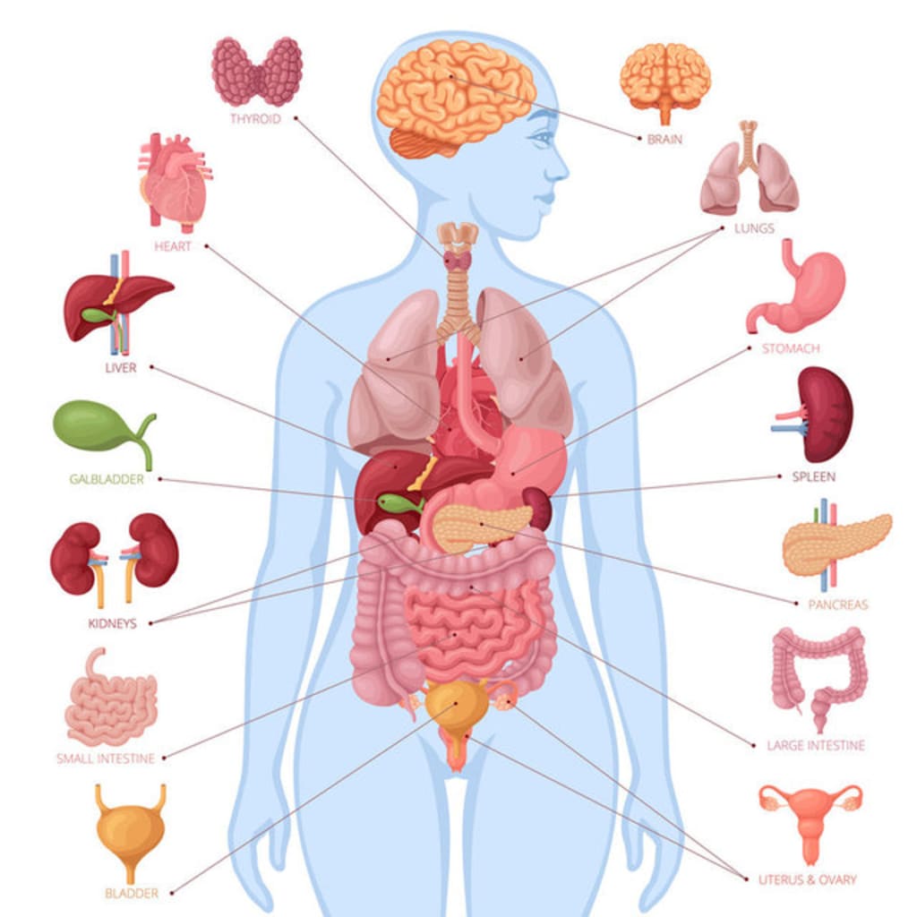
Accessory Spleen: How Many Spleens Do You Have?
The human body is a remarkable system consisting of various organs, each with its specific functions. While most people are familiar with major organs such as the heart, lungs, and liver, there are certain lesser-known structures that can also be present in the body. One such intriguing phenomenon is the accessory spleen, which raises the question: How many spleens do you have? In this article, we will explore the concept of accessory spleens, their significance, and their potential implications for human health.
Understanding the Accessory Spleen:
The spleen is a vital organ located in the upper-left side of the abdomen, known for its role in filtering the blood, removing old or damaged red blood cells, and assisting the immune system. However, in some individuals, small additional spleen-like structures, called accessory spleens, may be present. These structures are typically found in proximity to the main spleen but can occur in various locations within the abdomen.
Occurrence and Development:
The presence of accessory spleens is relatively common, with studies estimating that up to 10% of individuals may have them. These structures can vary in size, ranging from a few millimeters to several centimeters. The development of accessory spleens occurs during fetal development when certain clusters of cells fail to merge with the main spleen during organogenesis. Instead, they develop independently, resulting in the presence of multiple spleen-like structures.
Significance and Function:
While the main spleen is responsible for crucial functions related to blood filtration and immune response, the accessory spleens are generally considered to have limited or no functional significance. They are often inactive and do not actively participate in the filtering or immune processes performed by the primary spleen. However, in certain cases, if the main spleen is damaged or removed due to illness or surgery, accessory spleens may become more active and assume some of the functions of the primary spleen.
Diagnostic Considerations:
The presence of accessory spleens is typically asymptomatic and does not require any specific treatment or intervention. However, it is important to be aware of their existence, as they can be mistaken for other abdominal masses or tumors during imaging studies or surgical procedures. Accurate identification of accessory spleens can help prevent unnecessary investigations or potential complications during surgical interventions.
Conclusion:
The concept of accessory spleens adds an intriguing aspect to the understanding of human anatomy. While most individuals have a single spleen performing vital functions, the presence of accessory spleens highlights the remarkable diversity and variations that can occur in the human body. While these structures are generally considered to be of limited significance, their identification and differentiation from other abdominal masses are important for accurate diagnosis and appropriate medical management. If you have any concerns or questions regarding the presence of accessory spleens, consult with a healthcare professional for further evaluation and guidance.
"Superhuman" Olivia Farnworth: Never Hungry, Never Burned, Never Sleepy!
In the realm of extraordinary individuals, there are stories that captivate our imagination and challenge our understanding of human capabilities. One such remarkable person is Olivia Farnworth, often referred to as a "superhuman" due to her unique physiological traits. Olivia Farnworth defies conventional norms by exhibiting extraordinary characteristics such as never feeling hungry, never getting burned, and never experiencing the need for sleep. In this article, we delve into the fascinating world of Olivia Farnworth and explore the science behind her exceptional abilities.
The Enigma of Never Feeling Hungry:
One of the most astounding aspects of Olivia Farnworth's physiology is her apparent lack of hunger. While the majority of us experience regular sensations of hunger, Olivia claims to have never felt hungry in her entire life. This condition, known as congenital insensitivity to hunger, is an extremely rare genetic disorder that affects a minuscule fraction of the population. It results from a mutation in specific genes involved in regulating appetite and satiety, leading to a diminished or absent sense of hunger.
The Imperviousness to Burns:
Another extraordinary characteristic of Olivia Farnworth is her apparent immunity to burns. Despite being exposed to potentially hazardous situations, such as contact with hot surfaces or flames, she claims to never sustain burns or experience pain associated with them. This unusual ability can be attributed to a condition known as congenital insensitivity to pain with anhidrosis (CIPA). CIPA is an extremely rare disorder characterized by the inability to feel pain or perceive temperature changes due to abnormalities in the nervous system. While this condition provides Olivia with an apparent advantage in avoiding burns, it is important to note that it also poses significant challenges and risks, as pain serves as an essential protective mechanism.
The Mystery of Never Needing Sleep:
One of the most puzzling aspects of Olivia Farnworth's superhuman abilities is her claim of never needing sleep. Sleep is a fundamental physiological process crucial for rest, restoration, and overall well-being. However, Olivia states that she has never experienced the need for sleep and remains fully energized without it. While her case is exceptional and not fully understood, it is worth noting that a small subset of individuals, often referred to as "short sleepers," require significantly less sleep than the average person. This rare genetic trait allows them to function optimally on fewer hours of sleep without experiencing detrimental effects on their physical and cognitive abilities.
Implications and Scientific Exploration:
The extraordinary abilities exhibited by Olivia Farnworth open up intriguing avenues for scientific exploration and research. By studying individuals with congenital insensitivity to hunger, pain, or sleep, scientists can gain valuable insights into the underlying genetic and physiological mechanisms that govern these fundamental aspects of human existence. Such investigations may lead to breakthroughs in understanding appetite regulation, pain perception, and sleep-related disorders, potentially benefiting broader medical and scientific communities.
Conclusion:
Olivia Farnworth's extraordinary physiological traits of never feeling hungry, never getting burned, and never needing sleep capture our imagination and challenge our understanding of human capabilities. While her abilities may seem superhuman, they are grounded in rare genetic conditions that provide unique insights into the complexities of human physiology. Exploring and comprehending these exceptional cases not only expands our knowledge but also offers potential breakthroughs in medical research and the treatment of various conditions. Olivia Farnworth's story serves as a reminder of the remarkable diversity and potential of the human body and its ability to defy conventional limits.
Tetrachromats: Mutants Who Can See More Colors Than Us!
In the world of human perception, the ability to see colors is a remarkable gift. Most of us possess trichromatic vision, which means we can perceive and differentiate between thousands of colors using three types of color receptors in our eyes. However, there exists a small group of individuals known as tetrachromats, who possess an extraordinary ability to see a broader spectrum of colors than the average person. In this article, we delve into the fascinating world of tetrachromats, exploring the science behind their enhanced color vision and the unique experiences they encounter.
Unveiling the Mystery of Tetrachromacy:
Tetrachromacy is a rare condition characterized by the presence of an additional type of color receptor in the eyes, allowing for enhanced color discrimination. While the majority of humans have three types of color receptors, tetrachromats possess a fourth type, providing them with the ability to perceive a wider range of colors. This additional receptor, called the "cone," enables tetrachromats to distinguish subtle color variations that are imperceptible to trichromats.
The Genetics of Enhanced Color Vision:
The ability to be a tetrachromat is believed to be inherited through specific genetic mutations. It is passed down through generations, primarily through the X chromosome. Since males have only one X chromosome, the chances of them being tetrachromats are rare. However, females have two X chromosomes, increasing the likelihood of inheriting the mutation and potentially becoming tetrachromatic.
Exploring the Experience of Tetrachromats:
For tetrachromats, the world appears more vibrant and rich in color. They can discern subtle differences in hues, shades, and saturation that are imperceptible to trichromats. Colors that appear similar to the average person may appear distinct and separate to a tetrachromat. Their enhanced color vision offers a unique perspective on art, nature, and the surrounding environment.
The Challenges and Advantages of Tetrachromacy:
While tetrachromacy may seem like an extraordinary ability, it also comes with its challenges. The brain of a tetrachromat must process a vast amount of visual information, which can sometimes be overwhelming. Additionally, the rarity of tetrachromacy means that many individuals may not even be aware of their enhanced color vision, as they may perceive the world similarly to trichromats. However, those who are aware of their tetrachromatic abilities can develop a heightened appreciation for art, design, and visual aesthetics.
The Future of Tetrachromacy Research:
Tetrachromacy continues to be a subject of fascination for researchers and scientists. By studying individuals with tetrachromatic vision, scientists hope to unravel the intricate mechanisms behind their enhanced color perception. This research may lead to advancements in understanding color vision disorders, improving color technology, and expanding our knowledge of human visual perception.
Conclusion:
Tetrachromats possess a remarkable and rare ability to see a broader spectrum of colors than the average person. Their enhanced color vision opens up a world of vibrant hues and subtle variations that elude trichromats. Through genetics, biology, and the study of tetrachromats, we gain valuable insights into the complexity of human vision. Tetrachromacy serves as a reminder of the remarkable diversity of human perception and the potential for extraordinary sensory experiences.
ASD and PFO: Could Your Heart Have a Hole?
The human heart, a vital organ responsible for pumping blood throughout the body, is a marvel of biological engineering. However, there are certain conditions that can affect the structure and function of the heart, including the presence of holes in its chambers. In this article, we explore two specific conditions known as ASD (Atrial Septal Defect) and PFO (Patent Foramen Ovale) that involve a hole in the heart. We delve into the causes, symptoms, and potential treatments for these conditions, shedding light on the importance of early detection and appropriate medical care.
Understanding Atrial Septal Defect (ASD):
ASD is a congenital heart defect characterized by a hole in the septum, the wall that separates the two upper chambers of the heart, known as the atria. This hole allows oxygenated blood from the left atrium to mix with deoxygenated blood from the right atrium, leading to inefficient circulation. While small ASDs may not cause significant problems, larger defects can strain the heart and increase the risk of complications over time. Symptoms of ASD may vary, and some individuals may remain asymptomatic until later in life.
Exploring Patent Foramen Ovale (PFO):
PFO is another condition involving a hole in the heart, specifically in the septum between the two atria. However, unlike ASD, PFO refers to a persistent opening that should have closed shortly after birth. During fetal development, a small opening called the foramen ovale allows blood to bypass the lungs since they are not yet functional. In most individuals, this opening closes shortly after birth. However, in some cases, the closure may be incomplete, resulting in a PFO. Many individuals with a PFO may never experience any symptoms, while others may develop complications such as strokes or migraines.
Causes and Risk Factors:
Both ASD and PFO are congenital conditions, meaning they are present at birth. The exact causes of these conditions are not always known, but several factors may contribute to their development. Genetic predisposition, environmental influences, and certain medical conditions or syndromes can increase the risk of having a hole in the heart. Understanding these risk factors can help healthcare professionals identify individuals who may be at higher risk and provide appropriate monitoring and treatment.
Diagnosis and Treatment Options:
Diagnosing ASD and PFO often involves a combination of physical examinations, medical history reviews, imaging tests, and specialized procedures. Echocardiography, transesophageal echocardiography, and cardiac catheterization are commonly used to visualize and assess the presence and severity of the defects. Treatment options for ASD and PFO may vary depending on the size, location, and associated symptoms. Some small defects may not require immediate intervention but may be monitored over time. For larger or symptomatic defects, surgical procedures or minimally invasive techniques, such as catheter-based interventions, may be recommended to repair or close the holes in the heart.
Conclusion:
ASD and PFO are two conditions that involve a hole in the heart and can impact the proper functioning of this vital organ. Early detection, accurate diagnosis, and appropriate treatment are crucial for managing these conditions and minimizing the risk of complications. If you suspect you or someone you know may have a hole in the heart, it is important to consult with a healthcare professional for a comprehensive evaluation. Through medical advancements and increased awareness, we can enhance the care and well-being of individuals living with ASD and PFO, ultimately improving their heart health and overall quality of life.
Morton's Toe: Why Do Many Bulgarian Immigrants Have Unusual Toe Shape?
The human body is a remarkable creation, with each part serving a specific purpose. However, sometimes certain physical characteristics or traits can be more prevalent among specific populations or ethnic groups. In this article, we explore an intriguing phenomenon known as Morton's Toe, which is commonly found among Bulgarian immigrants. We delve into the causes, implications, and possible explanations for this unusual toe shape, shedding light on the intriguing connection between genetics, culture, and foot structure.
Understanding Morton's Toe:
Morton's Toe, also known as Morton's Foot or Greek Foot, refers to a condition where the second toe appears longer than the big toe. In a typical foot structure, the big toe is the longest, followed by the other toes in descending order. However, individuals with Morton's Toe have a longer second toe, which can sometimes extend beyond the length of the big toe. This unique toe shape can be observed in various populations worldwide, but it is particularly prevalent among Bulgarian immigrants.
Possible Explanations:
Genetic Factors: One possible explanation for the prevalence of Morton's Toe among Bulgarian immigrants lies in genetic factors. It is believed that certain genetic variations can contribute to the altered toe proportions. Studies have shown that the length of the toes is influenced by multiple genes, and the specific combination of these genes may be more common among Bulgarian individuals, resulting in a higher incidence of Morton's Toe.
Cultural Influence: Apart from genetic factors, cultural influences may also play a role in the prevalence of Morton's Toe among Bulgarian immigrants. Traditional footwear styles and cultural practices, such as wearing shoes with pointed or narrow toe boxes, can put pressure on the toes and contribute to the development of this toe shape. Over time, these cultural practices may have influenced the foot structure of Bulgarian individuals, leading to a higher occurrence of Morton's Toe.
Evolutionary Adaptation: Another fascinating perspective is that Morton's Toe may have conferred certain advantages in the past, providing evolutionary benefits to individuals with this foot structure. Some theories suggest that having a longer second toe could enhance balance, stability, or running capabilities. While further research is needed to support this hypothesis, it highlights the potential role of natural selection and adaptation in shaping the prevalence of Morton's Toe among specific populations.
Implications and Considerations:
Morton's Toe, in most cases, does not cause significant health issues or impair foot function. However, some individuals with this toe shape may experience discomfort, such as foot pain, calluses, or difficulties in finding properly fitting shoes. It is important to note that Morton's Toe is a natural variation and does not necessarily require medical intervention. For those experiencing discomfort, various solutions such as wearing shoes with wider toe boxes, using orthotic inserts, or seeking professional foot care can help alleviate any associated problems.
Conclusion:
The prevalence of Morton's Toe among Bulgarian immigrants is an intriguing phenomenon that intertwines genetic factors, cultural influences, and possibly evolutionary adaptations. Understanding the causes and implications of this unique toe shape adds to our appreciation of human diversity and the complex interplay between genetics and environment. While Morton's Toe is a distinctive characteristic, it does not pose significant health concerns for most individuals. Embracing and appreciating the diversity of human traits, including foot shapes, enriches our understanding of the complex tapestry of human biology and cultural heritage.
Anisocoria: Do Your Pupils Differ in Size?
The human body is a fascinating creation, full of unique features and functions. One intriguing phenomenon that can occur is anisocoria, a condition in which an individual's pupils have different sizes. In this article, we explore the causes, implications, and potential underlying reasons for anisocoria, shedding light on this fascinating aspect of human biology.
Understanding Anisocoria:
Anisocoria is a condition characterized by unequal pupil sizes. In most individuals, the pupils are relatively equal in size, allowing for balanced and coordinated vision. However, in some cases, one pupil may appear larger or smaller than the other. While anisocoria can be a natural variation, it can also be a symptom of an underlying medical condition.
Possible Causes:
Physiological Variation: In many cases, anisocoria is a benign and naturally occurring variation without any underlying medical conditions. The size difference between the pupils may be a result of differences in the muscle tone of the iris or variations in the nerve signals that control pupil size. These physiological differences do not typically cause any visual disturbances or health concerns.
Medications and Drugs: Anisocoria can also be a side effect of certain medications or drugs. Some substances, such as certain eye drops, medications for eye conditions, or even recreational drugs, can affect the muscles of the iris and result in unequal pupil sizes. If anisocoria develops after starting a new medication or drug, it is important to consult with a healthcare professional.
Underlying Medical Conditions: Anisocoria can sometimes be a symptom of an underlying medical condition. These conditions may include Horner's syndrome, a neurological disorder affecting the sympathetic nervous system, or Adie's tonic pupil, which is characterized by a larger pupil that reacts slowly to light. Other possible causes include brain injuries, tumors, or infections. If anisocoria is accompanied by other concerning symptoms, it is essential to seek medical evaluation.
Implications and Considerations:
In most cases, anisocoria is harmless and does not require medical intervention. However, it is crucial to be aware of any accompanying symptoms or changes in vision. If anisocoria is sudden, accompanied by pain, blurred vision, double vision, or other unusual symptoms, it is advisable to consult a healthcare professional for a comprehensive evaluation. They can determine the underlying cause and recommend appropriate treatment if necessary.
Conclusion:
Anisocoria, or unequal pupil sizes, can be a natural variation or a symptom of an underlying medical condition. While most cases of anisocoria are benign, it is important to pay attention to any accompanying symptoms or changes in vision. Regular eye examinations and open communication with healthcare professionals can help ensure early detection and appropriate management of any underlying conditions associated with anisocoria. Embracing the uniqueness of our bodies and seeking medical guidance when necessary allows us to better understand and care for the intricate workings of our visual system.
Double Jointedness: Is Having Hypermobile Joints "Normal"?
Have you ever come across someone who can bend their fingers, wrists, or limbs in seemingly impossible ways? Perhaps you have wondered if having such hypermobile joints is normal or a rare condition. In this article, we delve into the concept of double jointedness, scientifically known as joint hypermobility, to explore its characteristics, potential causes, and its impact on individuals.
Understanding Joint Hypermobility:
Joint hypermobility, commonly referred to as being "double jointed," is a condition characterized by unusually flexible joints. People with hypermobile joints have an extended range of motion, allowing them to move their limbs and body parts beyond the typical range most individuals can achieve. This increased flexibility can be observed in various joints, including the fingers, elbows, knees, and spine.
Causes and Factors:
The underlying causes of joint hypermobility can vary. In many cases, it is a genetic trait that is passed down through families. It is believed to be associated with differences in collagen, a protein responsible for providing structural support to the connective tissues in the body. Variations in collagen can affect the strength and stability of joints, leading to increased flexibility.
Additionally, certain medical conditions, such as Ehlers-Danlos syndrome and Marfan syndrome, are associated with joint hypermobility. These conditions involve abnormalities in connective tissues, which can result in joint laxity and hypermobility.
Is It Normal or Abnormal?
The classification of joint hypermobility as "normal" or "abnormal" is a topic of debate among medical professionals. While joint hypermobility itself is not considered a medical condition or a cause for concern, it can increase the risk of certain issues. Some individuals with hypermobile joints may experience joint pain, instability, or a higher susceptibility to joint injuries, such as dislocations or sprains.
Moreover, joint hypermobility can be associated with other symptoms or conditions, including chronic pain, fatigue, and joint-related disorders like osteoarthritis. In such cases, it is crucial for individuals to consult with healthcare professionals for proper evaluation and management of any associated symptoms or complications.
Management and Care:
For individuals with joint hypermobility, it is important to strike a balance between enjoying the benefits of flexibility and maintaining joint stability. This can be achieved through various means:
Regular Exercise and Strength Training: Engaging in exercises that focus on strengthening the muscles around the joints can help provide stability and support. This includes activities like resistance training, yoga, and Pilates.
Protective Measures: Taking precautions to avoid joint injuries, such as wearing appropriate footwear, using protective braces or supports, and practicing proper body mechanics during physical activities, can help minimize the risk of joint-related issues.
Seeking Professional Guidance: Consulting with healthcare professionals, such as physical therapists or orthopedic specialists, can provide personalized recommendations and guidance tailored to an individual's specific needs and concerns.
Conclusion:
Joint hypermobility, or being "double jointed," is a unique characteristic observed in some individuals. While it is not inherently abnormal or a medical condition, it is important to be aware of its potential implications and associated risks. By understanding the causes, implementing proper management strategies, and seeking professional guidance when needed, individuals with joint hypermobility can lead active and fulfilling lives while ensuring the well-being and long-term health of their joints. Embracing the diversity of our bodies and taking proactive steps for self-care are key to navigating the world of joint hypermobility.

About the Creator
Keep reading
More stories from Mert ÇANAKÇI and writers in Humans and other communities.
Secrets to Efficient Online Shopping: How to Shop Smart and Save
In today's digital age, online shopping has become increasingly popular. The convenience of being able to purchase desired products from the comfort of your own home while saving time and energy is unmatched. However, to make the most of your online shopping experience, it is essential to approach it with efficiency and smart decision-making. In this article, we will explore the secrets to efficient online shopping, providing you with valuable tips to shop smart and save.
By Mert ÇANAKÇI3 years ago in Trader
The CEO of Everything
They say "jack of all trades, master of none," but they forgot to mention the part where the jack of all trades is also the camera woman, the makeup artist, and the person currently yelling at a tangled Wii microphone cable in a her own bathroom while recording herself singing and trying to make it look realistic and professional. (As professional as you can make it with nothing but a cell phone camera and a mic that doesn't work)
By Sara Wilson7 days ago in Humans
Long Distance Relationship Questions That Test Real Love
Long-distance relationships are common, particularly among Gen Z couples in the United States. College migrations, career starts, visa concerns, and online contacts have all contributed to the prevalence of distance in modern love. Daily texting and video conversations do not determine if a long-distance relationship will endure. It is the questions we ask each other and the honesty with which we respond.
By Relationship Guideabout 15 hours ago in Humans
Affection and Healing for Yourself
During the night of the last quarter moon, I gathered my ritual supplies. I carefully handled the chunk of black tourmaline that would protect me from your overall negative and narcissistic energy. I carefully walked the house with my stick of selenite in hand, asking the universe to cleanse our working space.
By Alisha Wilkins ✒️🦋🖋️5 days ago in Fiction


Comments
There are no comments for this story
Be the first to respond and start the conversation.