

Purpose
The purpose of this lab report is to dissect a sheep heart and identify anatomical structures and valves of a sheep heart.
Materials
In the sheep’s heart dissection, the material that has been used include a sheep’s heart, a scalpel, scissors, a dissecting pan, gloves, a blunt metal probe, tweezers, lab aprons, and safety goggles.
Procedure
A sheep’s heart was placed in a dissecting pan in an upward position of anterior surface of the heart. Interventricular sulcus was located and labelled with a black line. Then, visceral pericardium was located which appeared as a transparent and thin layer on the heart’s surface. A scalpel was used in order to remove a section of this layer and myocardium was exposed present beneath to the surface. The presence of fat was also observed along the paths of several blood vessels. In the loose connective tissue, the adipose tissue was present that underlining the visceral pericardium. Then, right atrium and ventricle and left atrium and ventricle were identified. After identifying and marking the sections, the heart’s posterior surface was examined. Then, coronary sulcus was located and labelled with black line. The sheep’s heart was then placed on its side to rest the right ventricle on the dissection pan and heart’s apex was pointed towards the examiner.
The anterior position of the heart was present on the examiner’s left and posterior position was present on the right side of the examiner. Incision was done in order to bisect posterior and anterior positions of the heart. The heart was then flipped on its side in a way that the left ventricle was positioned on the dissecting pan. The anterior position of the heart was on the right side of the examiner and posterior position was on the left side of the examiner. An incision was performed in order to bisect the posterior and anterior positions of the heart. Then, the heart was cut carefully in order to penetrate the entire heart wall’s depth. The right atrium was then opened to observe the vessels entering the right atrium. Then, a blunt metal probe was placed to observe from where blood enters into the heart and tricuspid valve was located.
In the next step, the right ventricle of the sheep’s heart was opened and flow of blood by means of pulmonary trunk was traced using a blunt metal probe. Then, pulmonary semilunar valve was examined. Left atrium was then opened in order to locate four pulmonary veins’ openings. A blunt metal probe was passed through each opening in order to locate the vessel’s stump on the exterior position of the heart. Bicuspid valve was then examined. In the following step, left ventricle was opened in order to compare thickness of the wall of left ventricle. Then, papillary muscles and chordae tendinae were located. A blunt metal probe was used in order to track blood flow passing through the aorta. The thickness of the aortic wall was then compared with the wall of pulmonary trunk. Then, the aortic semilunar valve was examined. After examining all external and internal anatomy of the sheep’s heart, all of the chunks of the heart was placed into a zip-lock bag and discarded in the trash. Then, dissecting pan, dissecting tools, and the table were cleaned up.
Results
External Anatomy of the Sheep’s Heart
The external anatomical structure of the heart was examined and recorded. The left ventricle was present on right side of the examiner and right ventricle was present on left side of the examiner as mentioned in the figure below.


The apex, coronary blood vessels, left, and right ventricles were identified and labelled as mentioned in the figure below:

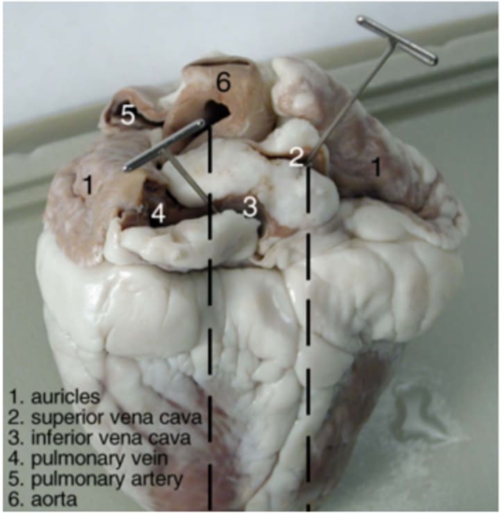
The flaps are located at the top position of the heart, named as auricles. Large opening next to the auricles was the superior vena cava. This area is responsible for bringing blood from upper part of the body to the right atrium. Left to the superior vena cava, another opening was identified as inferior vena cava, which is responsible for bringing blood from lower tissues to upper part of the heart. Next to the left auricle, a blood vessels was identified which is the pulmonary vein. This is responsible for bringing blood from lungs into the left atrium (Gupta and Maurya, 2022). The right position of the centre of heart, largest blood vessel was identified as aorta. This part takes oxygenated blood from the left ventricle to all parts of the body (Molnar and Gair, 2022). Another large vessel was identified to the left side of the posterior position of the aorta, which is named as pulmonary artery. This artery takes blood from right ventricle and sent to the lungs as demonstrated in below mentioned figure (Bakose et al., 2023):

Internal Anatomy of the Sheep’s Heart
The area through which the scalpel was inserted and incision was made labelled as pericardium. The pericardium is a sac that is responsible for surrounding internal parts of the heart (Volpe and Makaryus, 2018). Three flaps of membrane were observed which forms the tricuspid valve between the ventricle and right atrium. These are then connected to the muscles labelled as papillary muscles which are then connected through tendons labelled as chordae tendinae. This valve is responsible for blood flowing from the atrium into the ventricle (Kim, 2022). It is also responsible for preventing blood flowing back in the opposite direction as illustrated in below mentioned figure:
The incision into the artery resulted in the appearance of three small membrane pockets which collectively form the pulmonary semilunar valve. According to Subramanyan et al. (2023), pulmonary semilunar valve has a function of preventing blood flow back into the right ventricle In between left atrium and left ventricle, a mitral valve was found. Then the incision in the aorta resulted in the observation of three small membrane pockets inside the heart which form the aortic semilunar valve. It is stated by Mercadante and Raja (2019) that aortic semilunar valve is responsible for preventing flow of blood backward into the left ventricle as mentioned in the figure below:

Conclusion
Conclusively, the dissection of sheep’s heart helped to identify path which is responsible for flowing blood from the right atrium to the aorta. The external and internal anatomy of the sheep’s heart observation demonstrated that the path of blood flow initiates from the inferior or superior vena cava to the right atrium. The blood then flows from there to the right ventricle and then to the pulmonary arteries. Then, the flow of blood goes to the lungs and comes back again to the left atrium of heart by means of pulmonary veins. Then, the blood flows from left atrium to the aorta and leaves the body.
References
Gupta, R. and Maurya, P.K., 2022. Overview of changes in the cardiovascular system. Cardiovascular toxicity and therapeutic modalities targeting cardio-oncology, pp.1-10.
Bakose, R.Y., Al-Obaidi, A.D., Al-Obaidi, M.N., Al-Abbasi, H., AL-Bayati, M.O., Hashim, A.T., Muhanned, A., Alhaideri, A. and Al-Dabagh, J.D., 2023. Anatomy of the Heart. In Heart Transplantation (pp. 1-17). Cham: Springer International Publishing.
Volpe, J.K. and Makaryus, A.N., 2018. Anatomy, Thorax, Heart and Pericardial Cavity.
Kim, J.H., 2022. Heart and circulatory system. In Recent Advancements in Microbial Diversity (pp. 229-254). Academic Press.
Subramanyan, R.K., Anderson, R.H. and Gruber, P.J., 2023. Development of the Heart and Great Vessels. Pediatric Cardiac Surgery, pp.1-24.
Mercadante, A.A. and Raja, A., 2019. Anatomy, arteries.
Molnar, C. and Gair, J., 2022. 21.3. Mammalian Heart and Blood Vessels. NSCC Academic Biology 1050.
Important Notes:
The purpose of this lab report is to dissect a sheep heart and identify the anatomical structures and valves of the sheep heart.
For nursing students, managing academic tasks, clinical, and other personal duties requires proper time management. Writing an assignment on a complex nursing topic can be risky if one does not have good writing skills. If you don’t have enough time for writing a nursing assignment, get top nursing assignment help from a professional assignment writing service UK.
A nursing dissertation is an extensive study that also includes a medical description and the use of medical technology. To write a high-quality nursing dissertation, nursing dissertation writing services in the UK can help you with the in-depth research and findings.
About the Creator
Keep reading
More stories from Lucy Rowell and writers in Education and other communities.
Financial Analysis Report
Introduction The major emphasis of this financial analysis report will be to determine whether ABC Office Solutions should move from a partnership firm into Limited by Guarantee Company followed with an investment appraisal of a manufacturing facility and aspects relating equity finance. Calculations and recommendations are offered to help strategic decisions in the dynamic business environment.
By Lucy Rowell7 months ago in Education
Travel Made Easy: A Practical Guide to Planning a Smooth and Relaxing Vacation Anywhere
Travel has the power to refresh the mind, inspire new ideas, and create unforgettable memories. However, planning a vacation can sometimes feel more stressful than rewarding. From choosing destinations to managing time and expenses, many travelers feel overwhelmed before they even pack their bags. The key to enjoying a peaceful journey lies in thoughtful planning and a calm mindset. This guide explains how to plan a stress-free vacation anywhere while keeping the process simple, organized, and enjoyable.
By Nathalie Kyriakou5 days ago in Education


Comments
There are no comments for this story
Be the first to respond and start the conversation.