

“What do you do down there anyway?”
“What do pathologists do regarding surgical specimens?”
Let’s go back to a simpler time when I was a medical student, shall we? It was my second year in medical school and we were in the middle of a pathology rotation and I vaguely remembered it was a medical field containing very specialized consultants and soon became clear it was the field for me.
So quick story time before addressing the two questions I started this story with. (Also if bullet points bore you, this short story time will be where you leave me) Pathology was taught as a course in my 2nd year of medical school, but I also vaguely remembered it was a medical field containing very specialized consultants, an expert called in to look at things a different way and render a diagnosis or opinion. I decided that I needed to experience it first-hand.
There was a case scenario our professor wanted us to delve into. This case called upon us to look at a variety of tissues stained different shades of pink and blue and recognize specific features and patterns. When there are features we could not see, that is when those specialized consultants were called in. This type of consultant intrigued me. Who was this individual that knew more than us? What knowledge did he acquire that I did not have in order to diagnosis this tissue sample?
The importance of the pathologist’s diagnosis was astonishing; the course of treatment, along with the patient’s prognosis, was entirely dependent on the impression of the pathologist, the ultimate consultant. The field struck me as very cerebral; pathologists are required to have knowledge of virtually every disease process that can affect every organ in order to diagnose them.
Now fast forward through the rest of medical school and a few years into a Pathology residency and here we are today. A common theme upon the questions most pathologists may come across may often encompass the question of what is it we do with these specimens and samples sent to the lab.
This transition seems as good as any to let my readers whom dont enjoy a more expository writing style…well I bid you farewell.
There are four main "duties" that pathology residents have during surg path. At my institution, we cycle through these every few days, then start over.
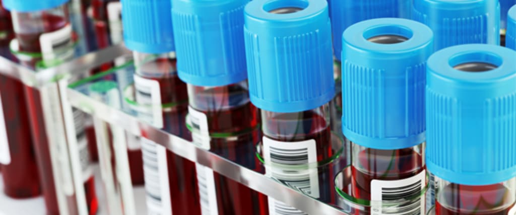
Frozen sections. Let's say a surgeon is dealing with a nasty invasive cancer. He cuts it out, but he wants to make sure he got all of it. He takes a margin (ie, a piece of tissue just past the edge of the tumor) and sends it to the frozen section room. There, the pathologist cuts the tissue up, freezes it with a special compound, uses a cryostat (a machine that holds the frozen tissue in place and cuts super-thin slices of it) to place a piece of the tissue on a slide, stains the slide with hematoxylin and eosin, then looks at it under the microscope to say whether there's cancer there. The slides this technique produces aren't quite as good as "permanent" slides (see below), but they'll do in a pinch. If there's cancer in the margin, the surgeon has to go back and take out more tissue. Frozen sections are done for various other reasons, including transplant viability (they want to put a fresh liver in a patient -- how healthy is it really?) The making of a frozen section isn't terribly difficult, but some tissues are hard to orient or cut properly, and when five surgeons send you specimens all at at once and wonder what's taking the lazy pathologist so long. It can get quite hectic.
2. Grossing. Anything that gets taken out during surgery -- a gallbladder, a cancerous kidney, a diabetic leg, a jaw full of tumor -- gets sent to us for grossing. We inspect the tissue, describe it, cut it open, and take representative sections. For the gallbladder, that means one section of mucosa (to see if it's inflamed) and one section of the cystic duct margin (to see if it's blocked). For the cancerous kidney, we take sections of the cancer (to see what kind of cancer it is) and sections around the cancer (to see if the cancer invaded the capsule / fat around the kidney / draining system of the kidney / renal vein / etc.).
This is a lot harder than it sounds, especially with weird specimens (the aforementioned jaw), complicated specimens (a Whipple -- ie, parts of stomach, pancreas, duodenum, and sometimes more), and annoying specimens (any colon cancer requires that we find ten lymph nodes in the fat -- which means mashing through the fat and hunting for tiny lymph nodes, usually for 90-120 minutes per colon). The last processor cuts off at 9 p.m., and that deadline is sometimes missed. The pieces of tissue removed from these specimens get placed in a processor and fixed in formalin overnight. The next day, they are turned into "permanent" slides in a more controlled and clean version of the "frozen section" procedure described above.
3. Previewing cases. Once the slides are out, the resident looks at them before signing out with the attending. A fourth-year resident can make most diagnoses and get all the paperwork in order without breaking a sweat. (Like myself) Me, when I was a first year though…I was just happy if I can tell it's a cancer, any cancer. We also have to look up clinical history on the patients, and sometimes we order special immunostains. These stains come in handy for diagnosis (is this skin lesion melanoma, order a S100 stain and see if it's positive) and/or prognosis.
And so fourth…no pun intended sine we’re onto the fourth point.
4. Signing out cases. This involves sitting with the attending, giving them whatever information they may need about the clinical history or the gross findings, and listening to them teach you why something is what it is. Usually somewhat painless, though each attending has his own style of doing things, and you're always preoccupied handling the small tasks you forgot to take care of while you were previewing the day before.
And that was basically it for me. It seems relatively simple when I see it written on my computer screen, but I must admit that the decision to become a pathologist was also in part an unscientific “gut feeling.” Quite simply, I was happiest when I was on Pathology, even if I couldn’t put my finger on why. I knew that I would find my niche.
About the Creator
Kranthi_Reddy
I hope this profile finds you well.
Keep reading
More stories from Kranthi_Reddy and writers in Longevity and other communities.
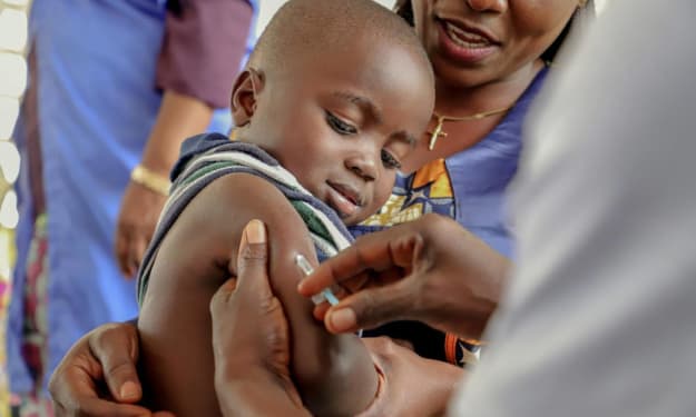
Development of Vaccinations: Within Child Medicine
Within the world of medicine, the search for vaccines has been on the forefront of the battle against a number of diseases that plagued humanity for centuries. Vaccines serve their purpose by preparing populations for the occurrence of certain diseases by introducing weakened or harmless forms of a virus in order for the body to acquire an immunity against its effects. Regardless of the era, vaccines remain a key part of public health, although there has been significant detraction from the ideas of vaccination due to perceived consequences of universally mandated vaccinations for children.
By Kranthi_Reddy4 years ago in Longevity
The Power of Black Coffee: Benefits, Nutrients, and Uses (Heath Tips)
Introduction: Black coffee is one of the most popular drinks in the world. Millions of people enjoy it every day, especially in the morning, because it helps them feel fresh, active, and focused.
By Health Tips6 days ago in Longevity
Chamomile Tea: Benefits, Nutrients, Best Time to Drink, and Complete Health Guide
Introduction: Chamomile tea is a popular herbal drink made from dried chamomile flowers. It has been used for centuries in traditional medicine because of its calming and soothing properties.
By Health Tipsabout an hour ago in Longevity


Comments
There are no comments for this story
Be the first to respond and start the conversation.