
3D Printing in Radiology: From Anatomical Models to Personalized Surgical Guides
3D Printing in Radiology

Introduction
The integration of 3D printing technology into the field of radiology has revolutionized medical imaging and surgical planning. No longer confined to the realm of science fiction, 3D printing offers tangible benefits to radiologists and surgeons alike, improving diagnostic accuracy, facilitating complex surgical procedures, and ultimately enhancing patient care. This transformative technology is bridging the gap between two-dimensional images and the intricate three-dimensional reality of the human anatomy, allowing for a level of precision and personalization previously unattainable. This article explores the expanding applications of 3D printing in radiology, from creating anatomical models for education and patient communication to the fabrication of highly customized surgical guides that improve surgical outcomes.
Anatomical Model Creation for Enhanced Understanding
The creation of accurate anatomical models from medical imaging data is a cornerstone of 3D printing's impact on radiology. These models provide radiologists with a more intuitive understanding of complex anatomical structures and pathologies. By holding a physical representation of a patient's unique anatomy, radiologists can better visualize the spatial relationships between different organs and tissues, facilitating more accurate diagnoses and treatment planning. This is particularly valuable in cases of congenital anomalies or intricate vascular networks, where traditional 2D imaging can be challenging to interpret fully. The tactile nature of these models improves understanding for both the radiologist and the patient, thereby promoting better communication and shared decision-making in the healthcare process.
Furthermore, these 3D-printed models serve as invaluable teaching tools for medical students and residents. They offer a hands-on learning experience that surpasses the limitations of textbooks and traditional anatomical models. The ability to examine intricate details, such as the branching patterns of blood vessels or the precise location of a tumor, provides a deeper understanding of human anatomy and pathology. This enhanced learning environment ultimately leads to better-trained medical professionals, who are better equipped to handle the complexities of modern medical practice.
Patient Education and Improved Communication
The use of 3D-printed anatomical models extends beyond the realm of medical professionals, providing significant benefits for patient education and improved communication. Patients often struggle to grasp complex medical information presented solely through two-dimensional images or abstract explanations. Presenting a tangible 3D model of their specific anatomy, highlighting the location of a tumor, the extent of a fracture, or the planned surgical approach, significantly enhances understanding and reduces patient anxiety. This increased transparency fosters trust between the patient and the healthcare team, leading to better adherence to treatment plans and improved overall patient outcomes. The models can also be used to explain the surgical process, allowing patients to visualize the steps involved and feel more in control of their own healthcare journey.
Beyond improving communication, these models aid in informed consent processes. By providing patients with a clear and concise representation of their condition and the proposed treatment, healthcare professionals can obtain more meaningful informed consent, ensuring patients are fully aware of the risks and benefits involved. This participatory approach to healthcare empowers patients and encourages active involvement in their own care.
Pre-surgical Planning and Surgical Guide Creation
The application of 3D printing in pre-surgical planning has significantly advanced surgical precision and efficiency. By leveraging medical images like CT scans and MRI scans, surgeons can create highly accurate 3D models of the patient's anatomy. These models enable meticulous pre-operative planning, allowing surgeons to simulate surgical procedures, identify potential challenges, and develop optimal surgical strategies. This virtual rehearsal dramatically reduces the risk of complications during surgery and contributes to faster recovery times for patients. The level of detail provided by these models is unparalleled, allowing for the precise measurement of anatomical structures, the identification of critical vascular structures, and the planning of precise incision sites.
Moreover, 3D printing allows for the creation of highly customized surgical guides. These guides are precisely designed and fabricated based on the patient's unique anatomy, providing surgeons with a real-time template during the operation. These guides aid in accurate placement of implants, improve the precision of bone cuts, and minimize the risk of damaging surrounding tissues. This enhanced precision translates to shorter operation times, reduced blood loss, and improved patient outcomes. The ability to personalize surgical guides for each patient represents a significant advancement in minimally invasive surgical techniques, making complex procedures safer and more effective.
Challenges and Future Directions
While the applications of 3D printing in radiology are undeniably promising, several challenges remain to be addressed. The cost of 3D printers and materials can be substantial, limiting accessibility in some healthcare settings. Furthermore, the technical expertise required to acquire, process, and utilize medical image data for 3D printing necessitates specialized training for radiologists and technicians. Standardization of protocols and quality control measures are also essential to ensure the reliability and accuracy of 3D-printed models and surgical guides.
Despite these challenges, the future of 3D printing in radiology is bright. Ongoing research is focused on developing more cost-effective materials, improving the speed and accuracy of printing processes, and expanding the range of applications. The integration of artificial intelligence and machine learning is expected to further refine the accuracy of 3D models and enhance the design of personalized surgical guides. The continued development and implementation of 3D printing technology in radiology will undoubtedly lead to further advancements in diagnostic accuracy, surgical precision, and ultimately, improved patient care.
Conclusion
The integration of 3D printing into radiology has profoundly impacted the field, offering tangible improvements in diagnosis, treatment planning, and surgical procedures. From creating anatomical models for enhanced understanding and patient education to facilitating the production of personalized surgical guides, 3D printing is reshaping the landscape of modern medical practice. While challenges remain, the ongoing advancements in this field promise even greater advancements in patient care, making 3D printing an indispensable tool for radiologists and surgeons alike. The future of this technology holds immense potential for further revolutionizing the diagnosis and treatment of a wide range of medical conditions.
About the Creator
Keep reading
More stories from Dr. Andrew Gomes and writers in 01 and other communities.
From Screen to Strategy: Radiology's Role in Treatment Planning
Introduction Radiology, the medical specialty focused on medical imaging, plays a pivotal role far beyond simple diagnosis. Its contribution extends deeply into the strategic planning of patient treatment across a wide spectrum of diseases. From the initial assessment to guiding minimally invasive procedures and monitoring therapeutic response, radiological data forms the bedrock upon which effective and precise treatment plans are built. This intricate interplay between image analysis and treatment strategy is crucial for optimizing patient outcomes and improving overall healthcare efficiency. Understanding this crucial connection is paramount for both clinicians and patients alike.
By Dr. Andrew Gomes6 months ago in 01
Next-Gen Conversational AI: How Natural Language Processing Services Drive Chatbot Success
Conversational AI has long since evolved past simple rule-based bots that followed fixed scripts. Today's chatbots manage complex questions, comprehend intent over the course of long conversations, respond with context and adjust to individual user behavior over time. This shift did not occur overnight. It is the result of steady advancements in Natural Language Processing and how businesses have applied it by using modern development practices.
By Fenil kasundra4 days ago in 01
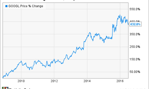
Goog Stock
Few companies command as much attention in the financial world as Google’s parent company, Alphabet Inc. Whether it’s innovation in artificial intelligence, shifts in digital advertising, or broader market volatility, Alphabet remains a central figure in global tech discussions. As a result, interest in Google stock—often searched as GOOG stock or GOOGL stock—tends to surge around earnings season, when investors look for clarity on growth, profitability, and long-term strategy. Alphabet operates with two publicly traded share classes: GOOG and GOOGL. While both represent ownership in the same company, GOOGL shares carry voting rights, whereas GOOG shares do not. Despite this distinction, the Alphabet stock price for both classes usually moves in near lockstep, reflecting the same underlying business performance and market sentiment. At the core of Alphabet’s valuation is Google’s advertising business. Search ads, YouTube ads, and display advertising across its network continue to generate the majority of revenue. During each Google earnings call, analysts closely examine advertising trends to assess whether businesses are increasing or cutting marketing budgets. Even modest changes in ad demand can significantly affect quarterly results, making Google earnings a key indicator for the broader tech sector. In recent quarters, Alphabet earnings have reflected a company navigating a complex environment. On one hand, Google maintains dominant market positions in search, mobile operating systems, and online video. On the other, it faces growing competition from social media platforms, e-commerce advertising, and emerging AI-driven tools. Investors tracking GOOG earnings and GOOGL earnings often focus on how well Alphabet balances efficiency with continued investment in innovation. Cloud computing has become another major factor shaping sentiment around Google stock. Google Cloud has grown steadily, narrowing losses and, in some periods, posting operating profits. While it still trails competitors in overall market share, progress in cloud services is seen as crucial to Alphabet’s long-term diversification. During each Google earnings call, executives are frequently asked about customer growth, enterprise adoption, and margins within the cloud division. Artificial intelligence has also moved to the center of the investment narrative. Alphabet has invested heavily in AI research for years, but recent developments have increased scrutiny on how those investments translate into revenue. From AI-powered search features to tools for advertisers and cloud customers, the company is under pressure to demonstrate that AI enhances profitability rather than simply increasing costs. Market reactions to Google earning reports often hinge on management’s guidance around AI-driven growth. Regulatory concerns remain a persistent backdrop. Alphabet faces antitrust scrutiny in multiple regions, including the United States and Europe. While these issues rarely impact quarterly earnings directly, they influence long-term investor confidence and can affect how analysts evaluate the Alphabet stock outlook. Legal expenses, potential fines, and changes to business practices are all factors that investors weigh when considering the sustainability of current profit margins. Another area investors monitor closely is capital allocation. Alphabet has used share buybacks to return value to shareholders, which can support the Alphabet stock price during periods of market uncertainty. Buybacks also signal management’s confidence in the company’s financial position. Combined with strong cash reserves, this approach has helped Alphabet maintain stability even during broader market downturns. Short-term movements in GOOG and GOOGL shares often reflect earnings surprises rather than long-term fundamentals. A stronger-than-expected advertising rebound or better cost control can push shares higher, while cautious guidance may lead to temporary pullbacks. For long-term investors, however, the focus tends to be on Alphabet’s ability to adapt to changes in how people search for information, consume content, and interact with digital services. The company’s “Other Bets” segment, which includes ventures like Waymo and life sciences initiatives, adds another layer of complexity. While these projects currently contribute little to revenue, they represent optionality that could drive future growth. Investors typically view them as long-term investments rather than immediate earnings drivers, but progress updates during Alphabet earnings reports can influence sentiment. Ultimately, Google stock reflects a balance between dominance and disruption. Alphabet remains one of the most profitable and influential companies in the world, yet it operates in an industry where technological shifts happen quickly. Each earnings report offers a snapshot of how well the company is navigating that reality. As markets continue to evolve, attention on GOOG stock and GOOGL stock is unlikely to fade. Whether investors are drawn by consistent cash flow, exposure to AI innovation, or long-term digital growth, Alphabet continues to occupy a central place in modern portfolios. For those watching closely, earnings season is more than a financial update—it is a window into how one of the world’s most powerful tech companies plans to shape the future.
By Saboor Brohi 3 days ago in 01


Comments
There are no comments for this story
Be the first to respond and start the conversation.